Tuesday, September 30, 2008

A DNA-based vaccine shows promise against avian flu

Posted: September 30, 2008

Though it has fallen from the headlines, a global pandemic caused by bird flu still has the United States’ Centers for Disease Control and Prevention on high alert. Yet, to date, the only vaccines that have proven even semi-effective are produced in chicken eggs, take five to six months to prepare and act against a single variant of the H5N1 virus, which mutates incredibly quickly. Now, new research by scientists in New York and Taiwan has led to a vaccine with the potential to stop most strains of H5N1 flu viruses in their tracks.

David D. Ho, Rockefeller’s Irene Diamond Professor and scientific director of the Aaron Diamond AIDS Research Center, together with his colleagues at Taiwan’s Academia Sinica, has built a vaccine that stimulates immunity to a broad range of H5N1 viruses in mice by using DNA rather than dead virus particles grown in eggs. Such a vaccine, which consists of plasmid DNA that’s been genetically modified to elicit specific immune responses, is much easier to rapidly modify and produce — critical advantages when racing to prevent an epidemic.

Ho and his collaborators first had to address virus specificity: Because H5N1 viruses are incredibly diverse, and mutate fast, the researchers created a consensus sequence that incorporated all of the conserved parts of the gene encoding the virus’s outer protein. Then they had to figure out how to deliver it.

This is where DNA vaccines often fail. They aren’t very good at making sure the DNA gets where it needs to go. To solve this problem, Ho and his colleagues turned to electroporation, a technique that is just beginning to gain traction in the vaccine world and that, according to preliminary studies, helps increase uptake of the vaccine. By combining their consensus-sequence vaccine with a small electric stimulus, the researchers found that their mouse subjects responded with an incredibly broad immune reaction.

“The immune responses directed to our DNA vaccine seem to be very broad,” Ho says. “It could be that the vaccine in its current form could protect against most of the H5N1 viruses out there.” And even if it can’t, he notes, if a different strain of H5N1 begins to circulate, it should only take a few days to obtain its genetic sequence and adapt the existing vaccine to fight it.

A version of the consensus vaccine is already being produced, Ho says, so that it can move into human clinical trials as quickly as possible. And a separate electroporation study is under way at The Rockefeller University Hospital, this one examining the effectiveness of electroporation combined with a DNA vaccine against HIV.

Proceedings of the National Academy of Sciences 105(36): 13538–13543 (September 9, 2008)

Contact: Zach Veilleux 212-327-8982
newswire@rockefeller.edu

Source
*
*

Sunday, September 28, 2008

Anticancer Drug Triggers Nanoparticle Formation, Improves Stability In Vivo

September 2008

Paclitaxel is a powerful anticancer agent used to treat a variety of malignancies, but severe side effects limit its ultimate effectiveness and utility. To improve this drug’s pharmacological properties, researchers have turned to nanoparticle-based delivery agents, although with marginal success; stably trapping large amounts of paclitaxel in most nanoparticles has proven difficult. Now, researchers at the University of Illinois at Urbana-Champaign have developed a new process for making nanoparticles that relies on paclitaxel itself to serve as the initiator that triggers polymer synthesis. The result is not only a stable nanoparticle formulation of paclitaxel but also one with very high and very controlled amounts of drug being incorporated in the nanoparticle.

Reporting their work in the journal Angewandte Chemie International Edition, Jianjun Cheng, Ph.D., a member of the Siteman Center of Cancer Nanotechnology Excellence (SCCNE), and graduate student Rong Tong, a student fellow funded by the SCCNE, describe the “living polymerization” technique they used to grow polymer nanoparticles that incorporate paclitaxel in the chemical structure of the polymer. The beauty of this process is that it provided the investigators with the ability to predetermine how much paclitaxel would end up in the nanoparticle merely by adjusting the ratio of drug molecule to lactide, the monomer from which polylactide is made. The investigators coat the final nanoparticle with a layer of poly(ethylene glycol) (PEG) to increase circulation time in the body.

Also important is the fact that the chemical linkage that binds paclitaxel to the polymer backbone can be broken down slowly in the body, providing sustained release of the drug once it reaches a tumor. Most nanoparticulate formulations of paclitaxel have been plagued by what researchers call “burst release,” which delivers as much as 90% of the drug within a few hours.

The researchers also note that this technique should be applicable for entrapping any drug that, like paclitaxel, contains hydroxyl groups in its chemical structure. In fact, they report that they have prepared stable nanoparticles containing the anticancer drugs docetaxel and camptothecin using their living polymerization technique.

This work, which is detailed in the paper “Paclitaxel-Initiated, Controlled Polymerization of Lactide for the Formulation of Polymeric Nanoparticulate Delivery Vehicles,” was supported by the NCI Alliance for Nanotechnology in Cancer, a comprehensive initiative designed to accelerate the application of nanotechnology to the prevention, diagnosis, and treatment of cancer. There is no abstract for this paper.


View journal citation

Source

PDF of Paper

Friday, September 26, 2008

A self-aligned single carbon nanotube field emission source fabricated by UV lithography

Click Pic To ENLARGE
Figure 5. SEM images of (a), (b) the gated carbon nanotube field emission source, where only one carbon nanotube grew per gate aperture
and was aligned at the center automatically, and (c) gate apertures with a 10 μm pitch.

Sewan Park et al 2008 Nanotechnology 19 445304 (7pp) doi: 10.1088/0957-4484/19/44/445304 Help


PDF (1.68 MB) | References


Sewan Park1,2, Hyeon Cheol Kim3, Min Hyung Yum4, Ji Hoon Yang4, Chong Yun Park4, Kukjin Chun1,2 and Bose Eom1,2
1 School of Electrical Engineering, Seoul National University, 599 Gwanangno, Gwanak-gu, Seoul 151-744, Republic of Korea
2 Inter-university Semiconductor Research Center, Seoul National University, 599 Gwanangno, Gwanak-gu, Seoul 151-742, Republic of Korea
3 School of Electrical Engineering, University of Ulsan, Daehakro 102, Nam-gu, Ulsan, 680-749, Republic of Korea
4 Department of Physics, Sungkyunkwan University, Chunchun-dong 300, Jangan-gu, Suwon, Gyeonggi-do, 440-746, Republic of Korea
E-mail: kchun@.snu.ac.kr and durian@mintlab.snu.ac.kr (K Chun)

Abstract. We suggest a novel process for fabricating a carbon nanotube field emission source having one carbon nanotube per gate aperture. The fabrication is based on UV lithography, instead of electron beam lithography. We used only one patterning step to define the gate, insulator, and cathode. We applied a DC voltage to the anode and a pulse signal to the gate. We then investigated the IV characteristics of the structure, changing the frequency and the duty-cycle of the pulse signal applied to the gate. We found that the optimum frequency and duty-cycle were 250 kHz and 22%, respectively. The structure had a turn-on voltage of 1.1 V under these conditions. The anode voltage did not have much effect. Finally, we checked the stability of the source for 40 h. We obtained an average emission current of 1.093 µA with a standard deviation of 1.019 × 10⁻² µA.

Print publication: Issue 44 (5 November 2008)
Received 17 June 2008, in final form 27 August 2008
Published 26 September 2008

Source

Graphene-stabilized copper nanoparticles as an air-stable substitute for silver and gold in low-cost ink-jet printable electronics

CLICK pic to ENLARGE

Figure 4. Printed copper patterns. (a) Line patterns for electrical
conductivity measurements. The inset shows a homogeneous and
crack-free print surface. (b) Manually cracked pattern for
investigation of the film thickness after five overprints, revealing a
print thickness of 500 nm per print cycle. The magnification inset of
the fracture surface shows the inner structure of the composite film.
(c) Graph showing the decrease of resistance with larger line width
of the pattern (error bars obtained from five samples at each line
width). The mean electrical conductivity was 1.56 ± 0.48 S cm−1.
(d) Light-emitting diodes directly glued onto printed copper lines.
Norman A Luechinger 2008 Nanotechnology doi: 10.1088/0957-4484/19/44/445201


PDF (611 KB) | References


Norman A Luechinger, Evagelos K Athanassiou and Wendelin J Stark1
Institute for Chemical and Bioengineering, Department of Chemistry and Applied Biosciences, ETH Zurich, CH-8093 Zurich, Switzerland
1 Author to whom any correspondence should be addressed
E-mail: wendelin.stark@chem.ethz.ch

Abstract. Metallic copper nanoparticles were synthesized by a bottom-up approach, and in situ coated with protective shells of graphene in order to get a metal nanopowder of high air stability and chemical inertness. Using an amphiphilic surfactant, a water-based copper nanocolloid could be prepared and successfully printed onto a polymer substrate by conventional ink-jet printing using household printers. The dried printed patterns exhibited strong metallic gloss and an electrical conductivity of >1 S cm-1 without the need for a sintering or densification step. This conductivity currently limits use in electronics to low current application or shielding and decorative effects. The high stability of graphene-coated copper nanoparticles makes them economically a most attractive alternative to silver or gold nanocolloids, and will strongly facilitate the industrial use of metal nanocolloids in consumer goods.

Print publication: Issue 44 (5 November 2008)
Received 11 July 2008, in final form 19 August 2008
Published 26 September 2008

Source

Thursday, September 25, 2008

Researchers Awarded Julius Springer Prize for Pioneering Work on Nanoscale Carbon Materials

This year's Julius Springer Prize for Applied Physics will be awarded to Phaedon Avouris and Tony Heinz for their pioneering work on the electrical and optical properties of nanoscale carbon materials including carbon nanotubes - from basic science to exciting applications. The award, accompanied by US$ 5,000, will be presented at the Julius Springer Forum on Applied Physics 2008 at Harvard University in Cambridge, MA, on 27 September 2008.

Future electronics and optoelectronics will be based on carbon nanostructures. Avouris and Heinz’s studies of the electronic properties of nanotubes and graphene aim at developing a future nanoelectronic technology with devices that will be vastly more compact, fast and energy efficient than the current silicon-based devices. The optoelectronic studies aim at uniting and integrating this electronic technology with an optical technology based on the same materials. Their research will aid in the development of future high-speed electronics, communications systems, and sensors for diverse applications. Industries ranging from automobile, aviation, space and energy conversion/conservation to bionanotechnology and medicine are likely to benefit from their research.

Phaedon Avouris received his B.Sc. degree from Aristotle University in Greece and was awarded his Ph.D. degree in physical chemistry at Michigan State University. He is currently an IBM Fellow and manager of Nanoscience and Nanotechnology at IBM’s Research Division at the Watson Research Center in Yorktown Heights, NY. He has also been an adjunct professor at Columbia University and the University of Illinois.

Tony Heinz earned his B.Sc. from Stanford University and his Ph.D. degree in physics from the University of California, Berkeley. He is the David M. Rickey Professor in the Departments of Physics and Electrical Engineering at Columbia University, where he has been since 1995. Previous to this, he worked at IBM’s Research Division at the Watson Research Center.

The Julius Springer Prize for Applied Physics recognizes researchers who have made an outstanding and innovative contribution to the fields of applied physics. It has been awarded annually since 1998 by the Editors-in-Chief of the Springer journals Applied Physics A – Materials Science & Processing and Applied Physics B – Lasers and Optics.

Springer is the second-largest publisher of journals in the science, technology, and medicine (STM) sector and the largest publisher of STM books. Springer is part of Springer Science+Business Media, one of the world’s leading suppliers of scientific and specialist literature.

Posted September 23rd, 2008

Source

New Nanoscale Process Created by UCSB Scientists Will Help Computers Run Faster and More Efficiently

Atomic Force Microscope image of a square array of 15nm pores formed by the new technology.
Abstract:
Smaller. Faster. More efficient. These are the qualities that drive science and industry to create new nanoscale structures that will help to speed up computers.

Scientists at the University of California, Santa Barbara have made a major contribution to this field by designing a new nanotechnology that will ultimately help make computers smaller, faster, and more efficient. The new process is described in today's Science Express, the online version of the journal Science.

Santa Barbara, CA | Posted on September 25th, 2008

For the first time, the UCSB scientists have created a way to make square, nanoscale, chemical patterns -- from the bottom up --that may be used in the manufacture of integrated circuit chips as early as 2011. It is called block co-polymer lithography.

Five leading manufacturers, including Intel and IBM, helped fund the research at UCSB, along with the National Science Foundation and other funders. The university has already applied for patents on the new methods developed here, and it will retain ownership.

A multidisciplinary team led by Craig Hawker, materials professor and director of the Materials Research Laboratory at UCSB, with professors Glenn Fredrickson and Edward J. Kramer, have developed a novel process for creating features on silicon wafers that are between five and 20 nanometers thick. (A nanometer is the thickness of one-thousandth of a human hair.)

Hawker explained that for the future we need more powerful microprocessors that use less energy. "If you can shrink all these things down, you get both," he said "You get power and energy efficiency in one package."

He said that the industry is up against Moore's law, a trend that Gordon Moore, Intel co-founder, first described in 1965 in which the power of the microprocessor doubles every 18 months. "One of the problems is that the industry is now running into physical limitations," said Hawker. "You can't shrink things down any more with the current technology." One of the ways that microprocessors are made is by using a top-down technique called photolithography, which involves shining light onto the surface of a silicon wafer, and making patterns. He explained that the size of the wavelength of light is becoming a limiting factor, and so his team has invented a new way of creating smaller patterns.

"We've come up with this new blending approach, called block co-polymer lithography, or BCP," said Hawker. "It essentially relies on a natural self-assembly process. Just like proteins in the body, these molecules come together and self assemble into a pattern. And so we use that pattern as our lithographic tool, to make patterns on the silicon wafer."

Using this technique, the size of the features is about the same as that of the molecules. They are very small, between five and 20 nanometers. "With this strategy, we can make many more features," said Hawker, "and hence we can pack the transistors closer together and everything else closer together --using this new form of lithography."

When this technique has been tried before, the molecules spontaneously self assembled into hexagonal arrays; they look like bee hives. But since industry uses parallel lines on a square or rectangular grid, the hexagonal arrays have limited application.

"In this article, we've actually shown that by changing the structure of the molecules, and using two self-assembling procedures at the same time, we're actually able to get square arrays, for the first time," said Hawker. "So now you can start to marry the old technology with the new technology for the fabrication of microprocessors."

Hawker said that the new technology was designed to be compatible with current manufacturing techniques, giving it the potential to be a "slip-in" technology. "All the big microprocessor companies like Intel and IBM have invested billions of dollars in their fabrication plants," said Hawker. "They're not going to throw out that technology anytime soon. It is too big of an investment and would not make good business sense. This allows them to introduce a new technology using current tools in the same fabrication plants. So they don't have to make huge up front investments to bring this to manufacturing. That's a key feature."

An analogy that Hawker uses in describing the development of the new methodology of block co-polymers is that of mixing salad dressing. "Think of the block co-polymers as oil and water," said Hawker. "When you make salad dressing you shake up the bottle because the oil and water don't want to be together. They separate into two layers. You shake your salad dressing and you mix everything up into much smaller droplets. What we've done is taken two polymer molecules that hate each other and joined them together. And so they want to separate just like the oil and water in your salad dressing. But because we've molecularly joined them, they can't. And so they separate into very, very small droplets, or domains, based on the fact that they hate each other. Those are the BCPs."

He explained that the interesting feature about this work is that the scientists combined the repulsive force with another self-assembly force which is slightly attractive.

"What we do is take one BCP (made of two components that hate each other) another BCP (again made of two components that hate each other) and simply mix these together," said Hawker. "When we mix them together, we've designed groups on one chain to be attracted to groups on a different chain, and so they actually start to blend and mix together. It is this combination of all these forces trying to get away from each other, and attract to each other that allows us to make the square arrays. Whereas what nature gives you is hexagonal, if you just use a single component system."

The scientists design the BCPs to have specific structures. And they use simulation to define the structures that are needed to prepare. "We design the molecule by understanding what needs to happen during the self-assembly process," said Hawker. "We need one block to be oil-like and one block to be water-like. So that's our first level of sophistication. We then design the molecular weight or the size of the molecule, to give us the desired feature size."

In the next step, the scientists design into the oil block the sticky groups that will form this attractive interaction, and by controlling the number of sticky groups, different levels of phase separation and different structures are created.

Polystyrene is the oil-like block, and one of the water-soluble blocks is polyethylene glycol. Polyethylene glycol is found in shampoos and many consumer products. It's a non-toxic, water-soluble, biocompatible polymer. By putting those together, the polyethylene glycol loves the water and the polystyrene loves the oil, and they hate each other. Polystyrene is found in disposable coffee cups, and according to the scientists is a fairly cheap commodity material that if designed in the right way, becomes a high value added application.

"The key to this work is that we put all the information into those molecules," said Hawker. "From a molecular level, we've built all the information into them that will allow them to undergo controlled phase separation. And the key is then just simply blending of two specifically designed materials, and then all we do is spin that down into a thin film on a silicon wafer. And then we heat it, and all the information that is pre-built into the molecule does its thing, and gives us the structure. And so that's why it is a really cheap technique. Because all you have to do is heat things up and you get the structures that you desire."

So the team has created a bottom-up approach to making these nanostructures, whereas the standard photolithographic technique, shining light onto the wafer -- is a top down engineering approach that requires multimillion dollar equipment.

In addition to Craig Hawker, the authors contributing the research, which was performed at UCSB, are: Chuanbing Tang, a postdoctoral fellow at the Materials Research Laboratory; Glenn Fredrickson, professor of chemical engineering and director of the Mitsubishi Chemical Center for Advanced Materials; Erin M. Lennon, a graduate student with Glenn Fredrickson at the time of the work; and Edward J. Kramer, professor of materials and of chemical engineering. (Lennon is now a National Science Foundation Research Training Group postdoctoral scholar at Northwestern University.)

####

For more information, please click here

Contacts:
Gail Gallessich
gail.g@ia.ucsb.edu
805-893-7220

FEATURED RESEARCHERS

Craig Hawker
hawker@mrl.ucsb.edu
805-893-7161

Glenn Fredrickson
ghf@mrml.ucsb.edu
805-893-8308

Edward Kramer
edkramer@mrl.ucsb.edu
805-893-4999

Copyright © University of California, Santa Barbara

Source

New Graphene-Based Material Clarifies Graphite Oxide Chemistry

September 25, 2008

AUSTIN, Texas — A new "graphene-based" material that helps solve the structure of graphite oxide and could lead to other potential discoveries of the one-atom thick substance called graphene, which has applications in nanoelectronics, energy storage and production, and transportation such as airplanes and cars, has been created by researchers at The University of Texas at Austin.

To get an idea of the nanomaterial graphene, imagine a lightweight material having the strongest chemical bond in nature and, thus, exceptional mechanical properties. In addition it conducts heat better than any other material and has charge carriers moving through it at a significant fraction of the speed of light. Just an atom thick, graphene consists of a "chickenwire" (or honeycomb) bonding arrangement of carbon atoms—also known as a single layer of graphite.

Mechanical Engineering Professor Rod Ruoff and his co-authors have, for the first time, prepared carbon-13 labeled graphite. They did this by first making graphite that had every "normal" carbon atom having the isotope carbon-12, which is magnetically inactive, replaced with carbon-13, which is magnetically active. They then converted that to carbon-13 labeled graphite oxide and used solid-state nuclear magnetic resonance to discern the detailed chemical structure of graphite oxide.

The work by Ruoff's team will appear in the Sept. 26 issue of the journal Science.

"As a result of our work published in Science, it will now be possible for scientists and engineers to create different types of graphene (by using carbon-13 labeled graphene as the starting material and doing further chemistry to it) and to study such graphene-based materials with solid-state nuclear magnetic resonance to obtain their detailed chemical structure," Ruoff says. "This includes situations such as where the graphene is mixed with a polymer and chemically bonded at critical locations to make remarkable polymer matrix composites; or embedded in glass or ceramic materials; or used in nanoelectronic components; or mixed with an electrolyte to provide superior supercapacitor or battery performance. If we don't know the chemistry in detail, we won't be able to optimize properties."

Graphene-based materials are a focus area of research at the university because they are expected to have applications for ultra-strong yet lightweight materials that could be used in automobiles and airplanes to improve fuel efficiency, the blades of wind turbines for improved generation of electrical power, as critical components in nanoelectronics that could have blazing speeds but very low power consumption, for electrical energy storage in batteries and supercapacitors to enable renewable energy production at a large scale and in transparent conductive films that will be used in solar cells and image display technology. In almost every application, sensitive chemical interactions with surrounding materials will play a central role in understanding and optimizing performance.

Ruoff and his team proved they had made such an isotopically labeled material from measurements by co-author Frank Stadermann of Washington University in St Louis. Stadermann used a special mass spectrometer typically used for measuring the isotope abundances of various elements that are in micrometeorites that have landed on Earth. Then, 100 percent carbon-13 labeled graphite was converted to 100 percent carbon-13 labeled graphite oxide, also a layered material but with some oxygen atoms attached to the graphene by chemical bonds.

Co-authors Yoshitaka Ishii and Medhat Shaibat of the University of Illinois-Chicago then used solid state nuclear magnetic resonance to help reveal the detailed chemical bonding network in graphite oxide. Ruoff says even though graphite oxide was first synthesized more than150 years ago the distribution of oxygen atoms has been debated even quite recently.

"The ability to control the isotopic labeling between carbon-12 and carbon-13 will lead to many other sorts of studies," says Ruoff, who holds the Cockrell Family Regents Chair in Engineering #7.

He collaborates on other graphene projects with other university scientists and engineers such as Allan MacDonald (Departments of Physics and Astronomy), Sanjay Banerjee, Emanuel Tutuc and Bhagawan Sahu (Department of Electrical and Computer Engineering) and Gyeong Hwang (Department of Chemical Engineering), and some of these collaborations include industrial partners such as Texas Instruments, IBM and others.

Co-authors on the Science article include: Weiwei Cai, Richard Piner, Sungjin Park, Dongxing Yang, Aruna Velamakanni, Meryl Stoller and Jinho An (all of the Ruoff research group at The University of Texas at Austin); Sung Jin An, formerly of Pohang University of Science and Technology (POSTECH-Korea) and a visiting graduate student in the Ruoff group during the study; Dongmin Chen (Beijing National Laboratory for Condensed Matter Physics, Institute of Physics, Chinese Academy of Sciences); Stadermann; and Ishii and Shaibat of the University of Illinois-Chicago.

A high-resolution photo of Ruoff is available. Learn more about Ruoff's work.

For more information, contact: Daniel Vargas, Cockrell School of Engineering, 512-471-7541; Rodney Ruoff, Department of Mechanical Engineering, Cockrell School of Engineering, 512-471-4691.

Source

Philips Announce Joint Research to Study Encapsulation of Magnetic Nanoparticles

Philips Research and the University of Urbino (Urbino, Italy) have signed a research agreement to study the encapsulation of magnetic nanoparticle contrast agents inside living blood cells to prolong the retention time of these agents in the blood.

Philips Research and the University of Urbino (Urbino, Italy) will jointly research new contrast agents for medical imaging that are based on the encapsulation of magnetic nanoparticles inside blood cells.

Injected as free particles, magnetic nanoparticle contrast agents are quickly excreted from the blood via the patient's liver, which limits their application. During the collaboration, the University of Urbino will investigate the integration of magnetic nanoparticles into red blood cells and their biological interactions in the human body, while Philips Research will evaluate the properties of these contrast agents in its medical scanners.

The collaboration between Philips Research and the University of Urbino will last for approximately two and a half years, with expected initial applications in the treatment of cardiovascular disease – one of the biggest killers in the western world.

"Nanoparticle blood pool contrast agents have already shown considerable promise in diagnostic imaging, but the short retention time of these particles in the body has always been a real challenge," says Henk van Houten, senior vice president of Philips Research and head of the Healthcare Research program. "Together with the unique expertise of the researchers at the University of Urbino we hope to increase the retention time of these particles from minutes to hours or even days, as this would open up applications such as the image-based monitoring of complex cardiovascular interventions that can take several hours to complete."

This healthcare research alliance follows the recently announced partnerships with West China Hospital in China, the University Medical Centers of Maastricht (the Netherlands) and Aachen (Germany), and the University Medical Center Utrecht in the Netherlands, and is part of Philips' increased commitment to developing solutions for improved patient care. A key success factor for this ambition is the effective translation from new concepts into clinical practice, which requires partnerships with leading academic and medical institutions. Bringing together such partnerships is one of the underlying principles behind Philips’ policy of open innovation.

“Our close collaboration with Philips should speed the translation of our invention into clinical practice,” comments Professor Mauro Magnani, Vice-Rector of the University of Urbino and a project leader of the EU FP6 funded NACBO (Novel and Improved Nanomaterials, Chemistries and Apparatus for Nano-Biotechnology) project. “With our technology, the use of new biomimetic constructs that merge the properties of nanomaterials with those of living cells is finally possible, bringing the real advantages of nanomaterials for therapeutic and diagnostic applications to patients.”

Posted September 24th, 2008

Source

Tuesday, September 23, 2008

Nanoparticle method may be able to inject drugs deep into the brain

Last Updated:10:01pm BST 22/09/2008

A new way to fight Alzheimer's and other neurological diseases aims to deliver a drug deep into the brain using nanoparticles

Alzheimer's is linked with the build up of damaging protein deposits in the brain and now a novel way to hinder this process is under development by a team at University College Dublin.

The team has a fundamental discovery in predicting how particles of the order of a billionth of a metre - nanoparticles - move around the body.

They report in the Proceedings of the National Academy of Sciences today that the size and the electrical charge of the nanoparticle matters, not just what it is made of.

The team found that a coating of proteins, called a corona, built up around the particles and this response mainly depends on the size and the charge of the nanoparticle.

This discovery, which shows that different sizes of the same materials pick up different proteins from the body, has a number of fundamental implications. "The find knocked my socks off," said Prof Kenneth Dawson, who led the work.

This find can be exploited to guide nanoparticles around the body.

The Dublin team is investigating how to exploit this to design nanoparticles to take an Alzheimer's drug to the brain.

Work by Prof Günter Oberdörster at the University of Rochester and Wolfgang Kreyling in Germany shows that nanoparticles can move into the brains of animals.

"It could change the face of health care if we learn how to exploit this potential to guide particles to important destinations in the body, such as the brain" said Prof Dawson.

"We have also now just found that certain nanoparticles are able to reverse the growth of Alzheimer plaques (the deposits of protein linked with the disease)," he said, explaining how they particles disrupt the smaller protein deposits that are thought to be the most toxic.

These experiments are only at the test tube stage, "but if we can combine the capacity of nanoparticles to get into the brain with this effect of reversing the growth of plaques that would point a way forward in these diseases."

By the same token of course, there should be caution and, as with any new technology, it must be "tested very carefully first," said Prof Dawson.

Thus, when people are exposed to nanoparticles they may penetrate into different parts of the body, with unknown health effects. So it is important to study the problems carefully, and not jump to conclusions, one way or the other, too quickly, he said.

Nanoparticles are as much as a million times smaller than the head of a pin, and have unusual properties compared with larger objects made from the same material.

The potential interactions of nanomaterials with the body and the environment have attracted increasing attention from the public as well as manufacturers of nanomaterial based products, academic researchers, and policy makers.

Nanoparticles can in principle make faster computers, and smaller mobile phones, as well as address some of the most intractable diseases. Nanotechnology is expected to become a $1 trillion industry within the next decade.

However, it is important that the be introduced safely and a new international research alliance to establish protocols for reproducible toxicological testing of nanomaterials in both cultured cells and animals was unveiled a few days ago at Nanotox 2008, a major research meeting.

This Alliance will be able to check each others work around the world, and build confidence in science, and further afield.

"When this team of scientists from Europe, the US and Japan are able to get the same results for interactions of nanomaterials with biological organisms, then science and society can have higher confidence in the safety of these materials," said Prof Dawson.

"This will open the doors to many potential benefits for society at large."

Source

Also see The Scientist: A new twist on nanoparticle behavior

Posted by Bob Grant
[Entry posted at 23rd September 2008 04:02 PM GMT]

Researchers hoping to develop nanoparticles as medicines or carriers of therapeutic molecules have much more to worry about than the type of material they plan on miniaturizing, according to a study in this week's issue of the Proceedings of the National Academy of Science.

Researchers in Ireland found that the corona, or cloud of proteins and other biomolecules that adheres to a nanoparticle immersed in biological media (in this study human blood plasma), changes depending on the size of the nanoparticle and the charge on its surface. That, in turn, can affect the particles' therapeutic action in the body.

Nanotechnology is "an enormously powerful tool, but we need to know how to control it," Kenneth Dawson, a University College Dublin physical chemist and the study's senior author, told The Scientist. "We have to look at what's happening at the surface of these materials rather than just the materials themselves. It's a new science really."

According to Dawson, the study represents a "paradigm shift" in how chemists typically think about the interaction of nanoparticles in biological settings. Traditionally the composition of the nanoparticle itself was thought to be the most important safety and functionality consideration. With Dawson's paper, the importance of the corona, and the physical factors which shape it, comes to the fore.

"The biological identity of a particle depends not only on its own material, but also what it picks up in the surroundings," Dawson said.

Dawson and his group last year coined the term "corona" for the conglomeration of proteins adhering to nanoparticles in biological media. Since then, the group has been exploring the properties of coronas. In the present study, the researchers found that nanoparticles introduced, in vitro, to human plasma accumulated markedly different coronas depending on their size and charge.

For example, his team found that uncharged particles attracted more immunoglobulins while charged particles pulled in more fat-shuttling proteins. "They pulled on quite different proteins," Dawson explained.

This, Dawson said, could have major implications for nanoparticles used as human therapeutics. For example, a particle of one size and surface charge might be trafficked to the brain of a patient, while another particle of a different size and charge, even though it's made of the same material, might be shuttled to the liver. "[A nanoparticle] can go places you didn't want it to go, and when it gets there it might pick up different signals that can be confusing," he said.

Drug makers and regulators should consider the effects of nanoparticle size and surface when developing and monitoring therapies that use nanotechnology, he added.

University of Rochester professor of environmental medicine and toxicology Gunter Oberdorster, who was not involved in the study, noted that drug makers may be able to make use of the differing physiological effects different coronas have on a nanoparticle's fate in the body. "Nanomedicine may take advantage of this and target a specific organ." While he called Dawson's study "an important step," he cautioned that in vivo studies must confirm the effects of nanoparticle size and surface character in living organisms.

According to Dawson, other researchers are conducting preliminary in vivo studies in mice to explore how nanoparticle sizes and surface properties affect the physiological activity of the tiny particles in living systems.

Although no treatments or therapeutics that use nanoparticles are currently on the market, experimental cancer treatments and other nanotechnology-based therapies are nearing FDA approval, according to Rice University chemist and Director of the International Council on Nanotechnology, Kristen Kulinowski. "This [study] bolsters the argument about the vital need for good, quality characterization [of nanomedicines], the actual species the body will experience," she told The Scientist.


Jefferson scientists deliver toxic genes to effectively kill pancreatic cancer cells

Public release date: 23-Sep-2008

Contact: Ed Federico
ed.federico@jefferson.edu
Thomas Jefferson University

New 'suicide gene' delivery approach offers potential for novel therapy

PHILADELPHIA – A research team, led by investigators at the Department of Surgery at Jefferson Medical College of Thomas Jefferson University and the Kimmel Cancer Center at Jefferson, has achieved a substantial "kill" of pancreatic cancer cells by using nanoparticles to successfully deliver a deadly diphtheria toxin gene. The findings – set to be published in the October issue of Cancer Biology & Therapy – reflect the first time this unique strategy has been tested in pancreatic cancer cells, and the success seen offers promise for future pre-clinical animal studies, and possibly, a new clinical approach.

The researchers found that delivery of a diphtheria toxin gene inhibited a basic function of pancreatic tumor cells by over 95 percent, resulting in significant cell death of pancreatic cancer cells six days after a single treatment. They also demonstrated that the treatment targets only pancreatic cancer cells and leaves normal cells alone, thus providing a potential 'therapeutic window.' Further, they are targeting a molecule that is found in over three-quarters of pancreatic cancer patients.

"For the pancreatic cancer world, this is very exciting," says the study's lead author, molecular biologist Jonathan Brody, Ph.D., assistant professor, Department of Surgery at Jefferson Medical College of Thomas Jefferson University, who works closely with the Samuel D. Gross Professor and Surgeon, Charles J. Yeo, M.D. "There are no effective targeted treatments for pancreatic cancer, aside from surgery for which only a minority of patients qualify. We are in great need of translating the plethora of molecular information we know about this disease to novel therapeutic ideas."

Pancreatic cancer is the fourth leading cause of cancer-related mortality in the U.S., reflecting the generally short survival time of patients - often less than a year from diagnosis.

This approach was originally developed in ovarian cancer cells by study co-author Janet Sawicki, Ph.D., a member of the Kimmel Cancer Center, and professor at the Lankenau Institute for Medical Research in Wynnewood, Pennsylvania. She and her group had recent success in reducing the size of ovarian tumors following treatment with diphtheria toxin nanoparticles.

The strategy is based on the fact that both ovarian and pancreatic cancer cells significantly over-express a protein found on the cell membrane, called mesothelin. The function of that molecule is unknown, but it is found in the majority of pancreatic tumors and ovarian cancer tumors. Other solid tumors also express mesothelin, but not at such a high rate.

"We don't know completely why cancer cells repeatedly turn on mesothelin genes to produce these membrane proteins, but it gives us a way to fool the cell and hijack its machinery, to trick it into making other more potent genes that will be detrimental to the cancer cells," Brody says.

To do that, the researchers devised an agent that consists of a bit of mesothelin DNA connected to the gene that produces the toxin from diphtheria, a highly contagious and potentially deadly bacteria, which is now controlled through childhood DPT vaccination. "Naked" DNA is then coated in a polymer to form nanoparticles that are taken up by the cancer cells.

Inside the cells, the agent performs its trickery. The nanoparticles biodegrade and the cell machinery senses genetic material from mesothelin. It activates the diphtheria toxin gene, which then turns on production of the toxin which allows the toxin to then do its work on the cancer cells, Brody says. Within 24 hours of delivery, the toxin disrupted production of protein machinery by over 95 percent, and within six days, a number of cancer cells die or are arrested.

"The cancer thinks it is turning on mesothelin and once it gets started reading that genetic code, it can't stop," he says. "So it will read the bacteria's DNA and produce the toxin which shuts down protein production in the cancer cells."

"It worked well in our cell culture models and now we are moving into pre-clinical experiments," Brody says.

The agent will not attack normal cells because the molecular machinery needed to turn on mesothelin is not found in normal cells, Brody says. Additionally, Sawicki has modified the diphtheria DNA to ensure that toxin that might be released from dying cancer cells is not taken up by healthy, normal cells.

But the researchers are now perfecting even more stringent measures to ensure safety, he says. "We can't help being hopeful," he says. "Our findings suggest that such a strategy will work in the clinical setting against the majority of pancreatic tumors."

Source

Saturday, September 20, 2008

Lack of vitamin D linked to multiple sclerosis

Saturday, September 20, 2008
Published: 7 hours ago

Children later diagnosed with multiple sclerosis had far lower levels of vitamin D than other youngsters, Canadian researchers reported yesterday in studies showing more links between the "sunshine" vitamin and disease.

These were the first studies to show the effects in children, although others have shown that adults who live in northern latitudes, who get less sun exposure, may have a higher risk of MS.

They also support a growing body of studies linking low vitamin D levels with disease, including cancer, heart disease, diabetes and tuberculosis.

Vitamin D, produced by the body when skin is exposed to sunlight, and also found in fatty fish, is added to other foods in many countries. Evidence suggests it helps lower blood pressure and boost the immune system.

Several studies presented at a meeting on MS in Montreal showed that children had low levels of vitamin D when they began to show evidence of the disease.

"Three-quarters of our subjects were below optimal levels for vitamin D," said Heather Hanwell, a graduate student in nutritional sciences at the University of Toronto, who led one study.

Hanwell's team studied 125 kids who had evidence of MS symptoms such as numbness. Twenty of the children were diagnosed with MS within the next year, Hanwell said. Blood tests showed 68 per cent of those children had vitamin D insufficiency.

On average, the children with MS had much lower levels of the vitamin than children who did not experience any other MS-like symptoms.

A study led by Brenda Banwell of Toronto's Hospital for Sick Children showed similar results.

"Seventeen of 19 children who had been diagnosed with MS had vitamin D levels below the target level," Banwell said.

The next step is to see if giving vitamin D supplements prevents MS or helps relieve symptoms, Banwell said. She said it was not clear how lacking vitamin D might be linked with MS.

Source

Friday, September 19, 2008

Pillared Graphene: A New 3-D Network Nanostructure for Enhanced Hydrogen Storage

ASAP Nano Lett., ASAP Article, 10.1021/nl801417w
Web Release Date: September 19, 2008

Copyright © 2008 American Chemical Society

Pillared Graphene: A New 3-D Network Nanostructure for Enhanced Hydrogen Storage

Georgios K. Dimitrakakis, Emmanuel Tylianakis, and George E. Froudakis*

Department of Chemistry, and Materials Science and Technology Department, University of Crete, P.O. Box 2208, 71003 Heraklion, Crete, Greece

Received May 16, 2008

Revised July 11, 2008

Abstract:
A multiscale theoretical approach was used to investigate hydrogen storage in a novel three-dimensional carbon nanostructure. This novel nanoporous material has by design tunable pore sizes and surface areas. Its interaction with hydrogen was studied thoroughly via ab initio and grand canonical Monte Carlo calculations. Our results show that, if this material is doped with lithium cations, it can store up to 41 g H2/L under ambient conditions, almost reaching the DOE volumetric requirement for mobile applications.


Source

Thursday, September 18, 2008

Seventh-grader shines with solar cell research

William Yuan won a $25,000 scholarship for his graduate level work

By Christina Lent
(news photo)

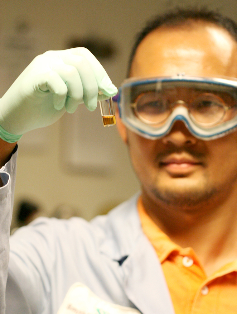
Jaime Valdez / The Beaverton Valley Times

William Yuan, 12, will be recognized Sept. 24 for his invention of a highly-efficient, three-dimensional nanotube solar cell for visible and ultraviolet light.

William Yuan’s bright idea to create a new, more efficient solar cell earned him top honors as Oregon’s only 2008 Davidson Fellow.

As part of the honor, the 12-year-old Bethany boy will be flown to Washington, D.C., for a reception Sept. 24 at the Library of Congress where he will receive his award and a $25,000 scholarship from the Davidson Institute for Talent Development.

“William’s work was evaluated by university professors and environmental scientists,” said Tacie Moessner, Davidson Fellows program manager in a call from Reno, Nev. “They look for the project’s potential to benefit society and make sure it is socially relevant. Generally, the projects need to be at the graduate level.”

Yuan worked on his project for the past two years with the encouragement of his science teacher Susan Duncan; support of his parents Gang Yuan and Zhiming Mei; and counsel of professional mentors Professor Chunfei Li of Portland State University’s Center for Nanofabrication and Electron Microscopy, Fred Li of Applied Materials Inc. and Professor Shaofan Li of the Department of Civil Engineering at the University of California – Berkeley.

“He is our youngest fellow in science that we’ve ever had,” Moessner said. “He is really spectacular.

“His project will really make a difference in advancing the technology of solar cells. You would never know he’s 12 looking at the quality of his work.”

Young talent

William Yuan is a seventh-grader in Meadow Park Middle School’s Summa options program.

He is an active member of the school’s Math Engineering Science Achievement (MESA) Club, First Lego League team and participant in the Science Bowl and MathCounts programs. He is also a two-time, second-place chess champion for the state.

Recognizing his interest in science, math and engineering, Yuan’s science teacher encouraged him to tackle a challenging engineering project for the Northwest Science Expo after introducing him to nanotechnology and renewable energy research.

“We learned about some great energy and environmental issues,” Yuan said. “To try to help, I researched the application of nanotechnology and renewable energy.

“I felt they would best complement my background knowledge and experience. After extensive research and community outreach, I wanted to work on a project to find a solution for some of the problems of the world.”

Yuan decided to focus his project on finding the most efficient way to harness the sun’s energy.

“I felt solar energy had large potential but it was underused,” he explained. “Fossil fuels like oil, coal and natural gas are only finite and are slated to run out by 2050.

“We need to make solar energy more cost effective and efficient.”

With that thought in mind, Yuan got to work.

“Current solar cells are flat and can only absorb visible light,” he said. “I came up with an innovative solar cell that absorbs both visible and UV light. My project focused on finding the optimum solar cell to further increase the light absorption and efficiency and design a nanotube for light-electricity conversion efficiency.”

Yuan invested countless hours in his research, seeking out new resources in the field to find a workable real-world solution.

“He has worked very hard in the past couple years,” his father Gang Yuan said. “We’re grateful that he had great mentors and teachers to guide him.

“When he started on his research, he had great curiosity and wanted to dig into it more. As his parents, we looked for experiences to help him.”

Watching his dedication impressed William’s parents.

“This generation’s sense of urgency is much stronger than my generation’s,” his father said. “They are thinking about the future and want to know how environmental issues will impact their generation.”

Promising future

Tapping into that talent and giving gifted youth the opportunity to excel is what the Davidson Institute is all about.

The national nonprofit organization recognized 20 students this year for their achievements.

Yuan admitted he submitted his project for review as a learning experience.

“This was a test run — I wasn’t expected anything,” he said. “I thought it would help when I entered another program when I was older.”

His work on developing his three-dimensional solar cell is far from complete.

“My next step is to talk to manufacturers to see if they will build a working prototype,” Yuan said. “If the design works in a real test stage, I want to find a company to manufacture and market it.”

The Davidson Institute scholarship will help Yuan further his research and his career in science and technology.

He plans to use the money to “attend one of the best universities in the country” and study nanotechnology, biotechnology or medicine.

“I’d like to work in technology at Google, Applied Materials or some other company that starts up between now and then,” Yuan said. “I’ve always liked math and science and engineering.

“If used properly, they can help solve the problems of the world. They can also be used to explore the world around us.”

Moessner has no doubt that Yuan will achieve great things in his future and looks forward to meeting him later this month.

“All of the fellows are really focused and driven but still humble,” she said. “They are also creative and brilliant.”

Source

*********************************************************************************

FWIW, this is the closest document I could find involving nanotubes, solar cells and ultraviolet, visible and infrared light:

United States Patent Application 20070240757
Kind Code A1
Ren; Zhifeng ; et al. October 18, 2007

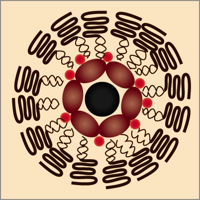
Solar cells using arrays of optical rectennas

Abstract

The present invention discloses a solar cell comprising a nanostructure array capable of accepting energy and producing electricity. In an embodiment, the solar cell comprises an at least one optical antenna having a geometric morphology capable of accepting energy. In addition, the cell comprises a rectifier having the optical antenna at a first end and engaging a substrate at a second end wherein the rectifier comprises the optical antenna engaged to a rectifying material (such as, a semiconductor). In addition, an embodiment of the solar cell comprises a metal layer wherein the metal layer surrounds a length of the rectifier, wherein the optical antenna accepts energy and converts the energy from AC to DC along the rectifier. Further, the invention provides various methods of efficiently and reliably producing such solar cells.


Inventors: Ren; Zhifeng; (Newton, MA) ; Kempa; Krzysztof; (Billerica, MA) ; Wang; Yang; (Allston, MA)


Assignee Name and Adress:
The Trustees of Boston College


Claims:
17. A method for producing a solar cell, comprising: growing a plurality of vertically-aligned nanotubes on a substrate; depositing a layer of a rectifying material onto the nanotubes; and depositing a layer of metal to cover a length of the nanotubes.

[0052] Aligned MWCNT arrays grown on silicon substrates using PECVD act as optical rectennas, receiving and transmitting light at ultraviolet (UV), visible and infrared (IR) frequencies.

Source

(WO/2008/112639) GRAPHITE-BASED PHOTOVOLTAIC CELLS

Pub. No.:
WO/2008/112639
International Application No.:


PCT/US2008/056420
Publication Date:18.09.2008 International Filing Date:10.03.2008
IPC: H01L 31/07 (2006.01), H01L 31/028 (2006.01), H01L 31/036 (2006.01)
Applicants:WISCONSIN ALUMNI RESEARCH FOUNDATION [US/US]; 614 Walnut Street, 13th Floor, Madison, Wisconsin 53726 (US) (All Except US).
LAGALLY, Max [US/US]; 5110 Juneau Road, Madison, Wisconsin 53705 (US) (US Only).
LIU, Feng [US/US]; 6681 South Candle Cove, Salt Lake City, Utah 84121 (US) (US Only).

SUMMARY OF THE INVENTION

[0006] The present invention encompasses graphite-based photovoltaic cells and methods for generating electricity from these cells. In these photovoltaic cells, spatially separated stacks of graphite, each comprising a plurality of vertically stacked semiconducting graphcnc(sic graphene) sheets, serve as a photovoltaic material bridging electrical contacts. The graphcnc(sic graphene) sheets, or "nanoribbons," have nanoscale-widtli(sic width) dimensions such that the band gap of each sheet depends on the width of the sheet. Thus, by incorporating graphenc(sic graphene) sheets having different widths, and thereby different band gaps, into the photovoltaic cell, the cell can be designed to absorb efficiently across the solar spectrum. The result is a photovoltaic cell that is efficient and inexpensive to manufacture.

Abstract:
The present invention uses lithographically patterned graphite stacks as the basic building elements of an efficient and economical photovoltaic cell. The basic design of the graphite-based photovoltaic cells includes a plurality of spatially separated graphite stacks, each comprising a plurality of vertically stacked, semiconducting graphene sheets (carbon nanoribbons) bridging electrically conductive contacts.


Source

Nano Carrier Targets Cell Sites

Thursday, September 18, 2008

Researchers find a new way to precisely target cancer drugs.

By Katherine Bourzac


Tiny target: A new targeted nano carrier selectively brings a cancer-killing drug to the mitochondria, the drug’s subcellular site of action. In these fluorescent images, yellow indicates that the drug is inside the mitochondria. Cell nuclei are stained blue.
Credit: Volkmar Weissig


Most drugs work by affecting a particular organelle within cells, but it's difficult to get a therapeutic compound to the right place inside a cell. Now researchers have succeeded in targeting a cancer-killing drug to a part of the cell called the mitochondrion by packaging it in a nano carrier. The highly targeted version of the drug increased its efficacy in tests in mice, even at relatively low doses, shrinking tumors and extending survival.

Over the past several years, researchers have had great success using antibodies and other molecules to target drugs to cells of particular tissue types. But once a drug gets inside the right cell, it's easy for it to get lost. Drugs are tiny compared with cells, and their charge, weight, and tendency to interact with water all determine where in the cell a drug ends up. "You have to design it such that it finds its way," says Volkmar Weissig, a professor of pharmacology at the Midwestern University College of Pharmacy, in Glendale, AZ, who developed the new targeted therapy with Vladimir Torchillin, director of the Center for Pharmaceutical Biotechnology and Nanomedicine, at Northeastern University, in Boston.

Subcellular targeting "is one of the biggest promises nanotechnology offers," says Jerry Lee, a project manager at the National Cancer Institute's Alliance for Nanotechnology in Cancer. The new research, he says, "offers early proof of concept of being able to target not only to cancer cells, but to pick and choose where in the cell to target."

Weissig and Torchillin developed a nano carrier to deliver a drug called ceramide to the mitochondria of cancer cells. The researchers enclosed ceramide within a sphere of lipids similar to those in many drug-delivery systems. This lipid envelope, which is too large to pass through the walls of healthy blood vessels, has a tendency to passively accumulate in tumors. (Tumor blood vessels have large gaps that allow the lipid-coated drugs in.) In order to actively target the drug to its subcellular site of activity, Weissig and Torchillin decorated the lipid envelopes with a molecule known to accumulate in the mitochondria.

In animal tests, the approach shows good efficacy, says Joseph DeSimone, a professor of chemistry and chemical engineering at the University of North Carolina at Chapel Hill. DeSimone is taking a different approach to intracellular targeting: he recently found that it's possible to control where in the cell nanoparticles accumulate by varying their shape. Overall, he says, "methods for accessing intracellular targets are extremely important to pursue."

Unhealthy mitochondria play a role in obesity and many diseases, including diabetes and degenerative diseases of the nervous system and muscle. And in theory, the nano-carrier system could be used to carry a wide variety of drugs to the mitochondria, says Weissig. However, since the carrier relies on leaky blood vessels to get to its target cells, it's not likely to be used to treat a wide variety of other diseases. Inflammatory diseases like arthritis, which also causes leaky blood vessels, are another possible application.

The nano-carrier technology was recently licensed by Telomolecular, a company in Rancho Cordoba, CA. Weissig says that the company will use it to develop an anticancer drug that works in the mitochondria. Although the system was proved using ceramide, Telomolecular will test other cancer drugs as well, says Weissig.

Source

Tuesday, September 16, 2008

New Carbon Material Shows Promise of Storing Large Quantities of Renewable Electrical Energy

September 16, 2008

AUSTIN, Texas — Engineers and scientists at The University of Texas at Austin have achieved a breakthrough in the use of a one-atom thick structure called "graphene" as a new carbon-based material for storing electrical charge in ultracapacitor devices, perhaps paving the way for the massive installation of renewable energies such as wind and solar power.

The researchers believe their breakthrough shows promise that graphene (a form of carbon) could eventually double the capacity of existing ultracapacitors, which are manufactured using an entirely different form of carbon.

"Through such a device, electrical charge can be rapidly stored on the graphene sheets, and released from them as well for the delivery of electrical current and, thus, electrical power," says Rod Ruoff, a mechanical engineering professor and a physical chemist. "There are reasons to think that the ability to store electrical charge can be about double that of current commercially used materials. We are working to see if that prediction will be borne out in the laboratory."

Two main methods exist to store electrical energy: in rechargeable batteries and in ultracapacitors which are becoming increasingly commercialized but are not yet as popularly known. An ultracapacitor can be used in a wide range of energy capture and storage applications and are used either by themselves as the primary power source or in combination with batteries or fuel cells. Some advantages of ultracapacitors over more traditional energy storage devices (such as batteries) include: higher power capability, longer life, a wider thermal operating range, lighter, more flexible packaging and lower maintenance, Ruoff says.

Ruoff and his team prepared chemically modified graphene material and, using several types of common electrolytes, have constructed and electrically tested graphene-based ultracapacitor cells. The amount of electrical charge stored per weight (called "specific capacitance") of the graphene material has already rivaled the values available in existing ultracapacitors, and modeling suggests the possibility of doubling the capacity.

"Our interest derives from the exceptional properties of these atom-thick and electrically conductive graphene sheets, because in principle all of the surface of this new carbon material can be in contact with the electrolyte," says Ruoff, who holds the Cockrell Family Regents Chair in Engineering #7. "Graphene's surface area of 2630 m2/gram (almost the area of a football field in about 1/500th of a pound of material) means that a greater number of positive or negative ions in the electrolyte can form a layer on the graphene sheets resulting in exceptional levels of stored charge."

The U.S. Department of Energy has said that an improved method for storage of electrical energy is one of the main challenges preventing the substantial installation of renewable energies such as wind and solar power. Storage is vital for times when the wind doesn't blow or the sun doesn't shine. During those times, the stored electrical energy can be delivered through the electrical grid as needed.

Ruoff's team includes graduate student Meryl Stoller and postdoctoral fellows Sungjin Park, Yanwu Zhu and Jinho An, all from the Mechanical Engineering Department and the Texas Materials Institute at the university. Their findings will be published in the Oct. 8 edition of Nano Letters. The article was posted on the journal's Web site this week.[See below]

This technology, Stoller says, has the promise of significantly improving the efficiency and performance of electric and hybrid cars, buses, trains and trams. Even everyday devices such as office copiers and cell phones benefit from the improved power delivery and long lifetimes of ultracapacitors.

Ruoff says significant implementation of wind farms for generation of electricity is occurring throughout the world and the United States, with Texas and California first and second in the generation of wind power.

According to the American Wind Energy Association, in 2007 wind power installation grew 45 percent in this country. Ruoff says if the energy production from wind turbine technology grew at 45 percent annually for the next 20 years, the total energy production (from wind alone) would almost equal the entire energy production of the world from all sources in 2007.

"While it is unlikely that such explosive installation and use of wind can continue at this growth rate for 20 years, one can see the possibilities, and also ponder the issues of scale," he says. "Electrical energy storage becomes a critical component when very large quantities of renewable electrical energy are being generated."

Funding and support was provided by the Texas Nanotechnology Research Superiority Initiative, The University of Texas at Austin and a Korea Research Foundation Grant for fellowship support for Dr. Park.

Learn more about Ruoff's work.

For more information, contact: Daniel Vargas, Cockrell School of Engineering, 512-471-7541; Rodney Ruoff, Department of Mechanical Engineering, Cockrell School of Engineering, 512-471-4691, 847-370-4637 (cell).

Source

Graphene-Based Ultracapacitors
Meryl D. Stoller, Sungjin Park, Yanwu Zhu, Jinho An, and Rodney S. Ruoff
Web Release Date: 13-Sep-2008; (Letter) DOI:
10.1021/nl802558y
Abstract Full: HTML / PDF (1703K)
Source

Researchers Develop Nano-Sized ‘Cargo Ships’ to Target and Destroy Tumors

September 11, 2008

By Kim McDonald

Scientists have developed nanometer-sized ‘cargo ships’ that can sail throughout the body via the bloodstream without immediate detection from the body’s immune radar system and ferry their cargo of anti-cancer drugs and markers into tumors that might otherwise go untreated or undetected.

Photo of Ji-Ho Park holding a vial containing the nanometer-sized cargo ships
UCSD graduate student Ji-Ho Park holds a vial containing the nanometer-sized cargo ships, composed of a magnetic nanoparticle, a fluorescent quantum dot and an anti-cancer drug molecule that will be left on the site of the tumor.

Credit: Luo Gu, UCSD

In a forthcoming issue of the Germany-based chemistry journal Angewandte Chemie, scientists at UC San Diego, UC Santa Barbara and MIT report that their nano-cargo-ship system integrates therapeutic and diagnostic functions into a single device that avoids rapid removal by the body’s natural immune system. Their paper is now accessible in an early online version here.

“The idea involves encapsulating imaging agents and drugs into a protective ‘mother ship’ that evades the natural processes that normally would remove these payloads if they were unprotected,” said Michael Sailor, a professor of chemistry and biochemistry at UCSD who headed the team of chemists, biologists and engineers that turned the fanciful concept into reality. “These mother ships are only 50 nanometers in diameter, or 1,000 times smaller than the diameter of a human hair, and are equipped with an array of molecules on their surfaces that enable them to find and penetrate tumor cells in the body.”

These microscopic cargo ships could one day provide the means to more effectively deliver toxic anti-cancer drugs to tumors in high concentrations without negatively impacting other parts of the body.

“Many drugs look promising in the laboratory, but fail in humans because they do not reach the diseased tissue in time or at concentrations high enough to be effective,” said Sangeeta Bhatia, a physician, bioengineer and professor of Health Sciences and Technology at MIT who played a key role in the development. “These drugs don’t have the capability to avoid the body’s natural defenses or to discriminate their intended targets from healthy tissues. In addition, we lack the tools to detect diseases such as cancer at the earliest stages of development, when therapies can be most effective.”

The researchers designed the hull of the ships to evade detection by constructing them of specially modified lipids--a primary component of the surface of natural cells. The lipids were modified in such a way as to enable them to circulate in the bloodstream for many hours before being eliminated. This was demonstrated by the researchers in a series of experiments with mice.

The researchers also designed the material of the hull to be strong enough to prevent accidental release of its cargo while circulating through the bloodstream. Tethered to the surface of the hull is a protein called F3, a molecule that sticks to cancer cells. Prepared in the laboratory of Erkki Ruoslahti, a cell biologist and professor at the Burnham Institute for Medical Research at UC Santa Barbara, F3 was engineered to specifically home in on tumor cell surfaces and then transport itself into their nuclei.

Photo of a vial of anti-cancer nano ships glows red under a black light.
A vial of anti-cancer nano ships glows red under a black light. The particles glow red because they contain fluorescent "quantum dot" nanoparticles.

Credit: Luo Gu, UCSD

“We are now constructing the next generation of smart tumor-targeting nanodevices,” said Ruoslahti. “We hope that these devices will improve the diagnostic imaging of cancer and allow pinpoint targeting of treatments into cancerous tumors.”

The researchers loaded their ships with three payloads before injecting them in the mice. Two types of nanoparticles, superparamagnetic iron oxide and fluorescent quantum dots, were placed in the ship’s cargo hold, along with the anti-cancer drug doxorubicin. The iron oxide nanoparticles allow the ships to show up in a Magnetic Resonance Imaging, or MRI, scan, while the quantum dots can be seen with another type of imaging tool, a fluorescence scanner.

“The fluorescence image provides higher resolution than MRI,” said Sailor. “One can imagine a surgeon identifying the specific location of a tumor in the body before surgery with an MRI scan, then using fluorescence imaging to find and remove all parts of the tumor during the operation.”

The team found to its surprise in its experiments that a single mother-ship can carry multiple iron oxide nanoparticles, which increases their brightness in the MRI image.

“The ability of these nanostructures to carry more than one superparamagnetic nanoparticle makes them easier to see by MRI, which should translate to earlier detection of smaller tumors,” said Sailor. “The fact that the ships can carry very dissimilar payloads—a magnetic nanoparticle, a fluorescent quantum dot, and a small molecule drug—was a real surprise.”

The researchers noted that the construction of so-called “hybrid nanosystems” that contain multiple different types of nanoparticles is being explored by several other research groups. While hybrids have been used for various laboratory applications outside of living systems, said Sailor, there are limited studies done in vivo, or within live organisms, particularly for cancer imaging and therapy.

“That’s because of the poor stability and short circulation times within the blood generally observed for these more complicated nanostructures,” he added. As a result, the latest study is unique in one important way.

Illustration of a vial of anti-cancer nano ships glows red under a black light.
The nanometer-sized cargo ships look individually like a chocolate-covered nut cluster, in which a biocompatible lipid forms the chocolate shell and magnetic nanoparticles, quantum dots and the drug doxorubicin are the nuts.

Credit: Ji-Ho Park, UCSD

“This study provides the first example of a single nanomaterial used for simultaneous drug delivery and multimode imaging of diseased tissue in a live animal,” said Ji-Ho Park, a graduate student in Sailor’s laboratory who was part of the team. Geoffrey von Maltzahn, a graduate student working in Bhatia’s laboratory, was also involved in the project, which was financed by a grant from the National Cancer Institute of the National Institutes of Health.

The nano mother ships look individually like a chocolate-covered nut cluster, in which a biocompatible lipid forms the chocolate shell and magnetic nanoparticles, quantum dots and the drug doxorubicin are the nuts. They sail through the bloodstream in groups that, under the electron microscope, look like small, broken strands of pearls.

The researchers are now working on developing ways to chemically treat the exteriors of the nano ships with specific chemical “zip codes,” that will allow them to be delivered to specific tumors, organs and other sites in the body.

Media Contact: Kim McDonald, 858-534-7572
Comment: Michael Sailor, 858-534-8188

Source