NanoViricides has proven it works on viruses so let's see it work on bacteria next!
November 6, 2017
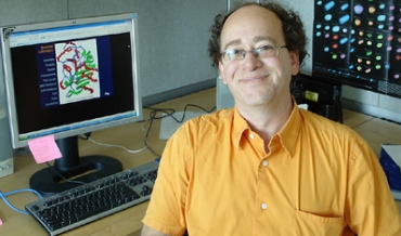
In the battle against drug-resistant bacteria, Marcos Pires studies the chemical biology of bacterial cell surfaces to better understand how they function -- and possibly how to manipulate them Credit: Hvass & Hannibal courtesy of Lehigh University
Cell walls—the jacket-like structures that surround all known bacteria—may turn out to be bacteria's undoing , holding the key to developing new drugs that target it for destruction.
That perspective is shared by many in the medical and scientific communities, including Marcos Pires . Pires, a biochemist at Lehigh University, is spearheading a novel approach to understanding bacterial cell wall changes in response to antibiotics that could be critical to new drug design—an urgent need in light of the growing threat of antibiotic resistance. His approach is so promising it has recently been recognized by the National Institutes of Health with a Maximizing Investigators' Research Award (MIRA).
Antibiotic resistance occurs when bacterial cells adapt to evade a drug designed to kill it. Making changes to the cell wall is one way bacteria accomplish this. Little is known, however, about just how these structures respond when under attack.
With the 5-year $1.94 million MIRA grant, Pires's group will delve deeply into this process through a unique approach that essentially tricks bacteria into revealing where its cell wall is most vulnerable. Such knowledge could help scientists design next-generation antibiotics that circumvent drug resistance mechanisms.
The centerpiece of the research is a process that Pires and his team conduct facilitating live bacteria's absorption of synthetic cell wall fragments constructed in the lab. These fragments are modified with reporter units which then allow researchers to observe, in live bacteria, components of the cell wall machinery under various conditions.
"Bacterial cell walls are unique in their structure and function and are essential to bacterial cells—making them unique targets for the development of antibiotics," said Pires, assistant professor in the Department of Chemistry. "By 'tricking' bacteria into using some of our cell wall building blocks, we get an unprecedented perspective on how they change when challenged with antibiotics."
MIRA is a program of the National Institute of General Medical Science (NIGMS), a division of NIH that provides support for basic research that increases understanding of biological processes and lays the foundation for advances in disease diagnosis, treatment and prevention. According NIGMS, the goal of MIRA is to increase the efficiency of NIGMS funding by providing investigators with greater stability and flexibility, thereby enhancing scientific productivity and the chances for important breakthroughs.
Identifying bacterial cell wall changes that cause antibiotic resistance
The stakes for drug design breakthroughs to treat drug-resistant bacteria are high. Every year in the United States, more than 2 million people are afflicted with resistant bacterial infections. An estimated 23,000 American lives—and 700,000 lives worldwide—are lost yearly as a result of bacterial infections resistant to current antibiotic treatments. These numbers are only expected to grow.
Bacterial cell walls are the target of some of the most powerful antibiotics discovered to date. Cell wall-targeting antibiotics include some commonly prescribed treatments such as penicillin and amoxicillin. Drugs that target bacteria's cell walls are also among the safest as human cells do not have cell walls and are thus unaffected by the treatment.
According to Pires, individual components of the bacterial cell wall machinery are key to bacteria's adaptation response and, therefore, to drug-resistance. One of his team's goals is to identify the cell wall components that bacteria need to successfully adapt and evade the drugs designed to destroy it.
"If we can identify these 'weak spots', said Pires, "we should be able to find ways to inactivate or circumvent them."
I'd send you a PM but you have me blocked. And as a non-paying member of iHub I don't have access to some features.
Anyway, wgas!
Ref:
Tristan V. Stonger - 2 Million PYHH
8/14/15 1,000,000 Common Stock N/A Tristan V. Stonger MD Marketing Services
8/18/15 1,000,000 Common Stock N/A Tristan V. Stonger MD Marketing Services
Of particular interest: William Friedman; Mark Stapp
These two individuals post often on iHub message boards and do NOT identify themselves as paid promoters as required. They received shares for their services - 50,000 each as noted.
iHub and SEC rules and regulations require that these sort of promotions be advertised - they have not been.
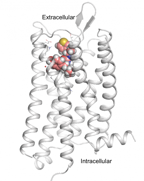
An international team of researchers — led by scientists at UC San Francisco, Stanford University, the University of North Carolina (UNC), and the Friedrich-Alexander University Erlangen-Nürnberg in Germany — has developed a new opioid drug candidate that blocks pain without triggering the dangerous side effects of current prescription painkillers. Their secret? Starting from scratch — with computational techniques that let them explore more than four trillion different chemical interactions.
In a new study — published online Aug. 17, 2016, in Nature — the researchers used the newly deciphered atomic structure of the brain’s “morphine receptor” to custom-engineer a novel drug candidate that blocked pain as effectively as morphine in mouse experiments, but did not share the potentially deadly side effects typical of opioid drugs. In particular, the new drug did not interfere with breathing — the main cause of death in overdoses of prescription painkillers as well as street narcotics like heroin — or cause constipation, another common opioid side effect. The new drug also appears to side-step the brain’s dopamine-driven addiction circuitry and did not cause drug-seeking behavior in mice.
More work is needed to establish that the newly formulated compound is truly non-addictive and to confirm that it is as safe and effective in humans as it is in rodents, the authors say. But if the findings are borne out, they could transform the fight against the ongoing epidemic of prescription painkiller addiction.
Deaths from opioid drug overdoses have been on the rise in the US for decades. According to the Centers for Disease Control and Prevention, 28,000 Americans died of narcotic overdoses in 2014, four times more than in 1999, with more than half of these deaths involving prescription drugs. The epidemic has gotten the attention of national leaders: in February, 2016, President Obama proposed $1.1 billion in new funding for opioid addiction treatment, and in July Congress passed the Comprehensive Addiction and Recovery Act, a bill intended to curb opioid abuse and improve treatment.
But as damaging as opioids can be, modern medicine depends on these drugs as our most powerful weapon against pain.
“Morphine transformed medicine,” said Brian Shoichet, PhD, a professor of pharmaceutical chemistry in UCSF’s School of Pharmacy and co-senior author on the new paper. “There are so many medical procedures we can do now because we know we can control the pain afterwards. But it’s obviously dangerous too. People have been searching for a safer replacement for standard opioids for decades.”
Virtual Experiments Lead to Novel Opioid Chemistry
Much of drug discovery, Shoichet says, begins by taking a successful drug like morphine and tweaking its structure to try to get rid of side effects while maintaining its primary function. The new study took a different, much more radical approach: “We didn’t want to just optimize chemistry that already existed,” Shoichet said. “We wanted to get new chemistry that would confer completely new biology.”
PZM21, the new, safer opioid drug candidate, is shown docked on the brain’s morphine receptor, the mu-opioid receptor. Image by Anat Levit
Key to the new paper was knowing the atomic structure of the mu-opioid receptor, the brain’s “morphine receptor,” which was recently deciphered by co-senior author and 2012 Nobel laureate Brian Kobilka, MD, a professor of molecular and cellular physiology at the Stanford University School of Medicine.
“With traditional forms of drug discovery, you’re locked into a little chemical box,” Shoichet said. “But when you start with the structure of the receptor you want to target, you can throw all those constraints away. You’re empowered to imagine all sorts of things that you couldn’t even think about before.”
With this structural information in hand, Shoichet’s team turned to a computational approach called molecular docking, which was pioneered in the 1980s at UCSF’s School of Pharmacy by Shoichet’s mentor, emeritus professor Tack Kuntz, PhD. In a two-week period, the researchers performed roughly four trillion “virtual experiments” on a UCSF computer cluster, simulating how millions of different candidate drugs could turn and twist in millions of different angles to find those configurations that were most likely to fit into a pocket on the receptor and activate it. They also strove to avoid molecules that could stimulate beta-arrestin2, part of a biological pathway linked to the respiratory suppression and constipation typical of other opioids.
This led to a short-list of 23 candidate molecules judged by the software and the research team — especially co-lead authors Henry Lin, PhD of UCSF and Aashish Manglik, MD, PhD at Stanford — to be most likely to activate the mu-opioid receptor in the way the researchers wanted.
Only then did the team actually test these candidate drugs in the real world. Co-lead author Dipendra Aryal, PhD, led a team of researchers in the pharmacology lab of co-senior author Bryan Roth, MD, PhD, a professor of pharmacology at the University of North Carolina (UNC) School of Medicine, to identify the most potent of the 23 leading candidates. Then, based on the structural insights of Manglik and Lin, Roth’s team worked with the lab of co-senior author Peter Gmeiner, PhD, chair and professor of medicinal chemistry at the Friedrich-Alexander University Erlangen-Nürnberg in Germany, to optimize this compound’s chemical efficacy 1000-fold. This approach succeeded in producing a molecule that the researchers called PZM21, which is chemically unrelated to existing opioid drugs.
‘Unprecedented, Weird and Cool’ New Biology
In further pharmacological tests conducted in the Roth lab, PZM21 exhibited the “new biology” the researchers had been looking for: efficiently blocking pain without producing the constipation and breathing suppression typical of traditional opioids. In addition, PZM21 appeared to dull pain by affecting opioid circuits in the brain only, with little effect the on opioid receptors in the spinal cord that mediate pain reflexes. No other opioid has such a specific effect, Shoichet said, calling it “unprecedented, weird and cool.”
Additional behavioral tests in mice suggested the drug may also lack the addictive qualities of existing opioids. Specifically, the drug didn’t produce the hyperactivity other opioids trigger in mice by activating the brain’s dopamine systems — which are also involved in addiction. Perhaps more tellingly, mice did not spend more time in test chambers where they had previously received doses of PZM21 — a test called “conditioned place preference” that is considered a correlate of human drug-seeking behavior.
Brian Shoichet, PhD
“We haven’t shown this is truly non-addictive,” Shoichet cautioned, emphasizing that further experiments in rats and humans would be needed to establish the compound’s addictive potential. “At this point we’ve just shown that mice don’t appear motivated to seek out the drug.”
The study is a successful example of the structure-based approach to drug discovery, a technique partially pioneered at UCSF 30 years ago, Shoichet said, and is one of the first to use structural knowledge to create fundamentally new biological effects.
“This promising drug candidate was identified through an intensively cross-disciplinary, cross-continental combination of computer-based drug screening, medicinal chemistry, intuition and extensive preclinical testing,” Kobilka said.
“If you took away any one of these collaborators it simply wouldn’t have worked,” Shoichet added. “Without Kobilka’s structure, our computation, Roth’s pharmacology, and Gmeiner’s ability to put an atom in exactly the place you want it, this never would have been possible.”
Lead authors on the new paper were Aashish Manglik, MD, PhD, of Stanford University School of Medicine; Henry Lin, PhD, of the UCSF School of Pharmacy; and Dipendra K. Aryal, PhD, of the UNC School of Medicine. Lin is now principal scientist at The Janssen Pharmaceutical Companies, a division of Johnson & Johnson. Manglik, Lin, Gmeiner, Kobilka, Roth, Shoichet, and co-author Dengler have filed a provisional patent on PZM21 and related molecules, and Manglik, Gmeiner, Kobilka, Roth and Shoichet are consultants and co-founders of Epiodyne, a company seeking to develop novel analgesics.
The research was supported by the US National Institutes of Health grants GM106990 (B.K.K., B.K.S. and P.G.), DA036246 (B.K.K.), GM59957 (B.K.S.), and the National Institutes of Mental Health Psychoactive Drug Screening Program (B.L.R.) and DA017204 (B.L.R., D.A.), DA035764 (B.L.R.) and the Michael Hooker Distinguished Professorship (B.L.R.) and the German Research Foundation Grants Gm 13/10 and GRK 1910 (P.G). H.L. received a pre-doctoral fellowship from the PhRMA Foundation and A.M. received support from the Stanford University Medical Scientist Training Program (T32GM007365) and the American Heart Association (12PRE8120001).
UCSF is a leading university dedicated to promoting health worldwide through advanced biomedical research, graduate-level education in the life sciences and health professions, and excellence in patient care. It includes top-ranked graduate schools of dentistry, medicine, nursing and pharmacy; a graduate division with nationally renowned programs in basic, biomedical, translational and population sciences; and a preeminent biomedical research enterprise. It also includes UCSF Health, which comprises two top-ranked hospitals, UCSF Medical Center and UCSF Benioff Children’s Hospital San Francisco, and other partner and affiliated hospitals and healthcare providers throughout the Bay Area.
Authors Li X, Ye X, Qi J, Fan R, Gao X, Wu Y, Zhou L, Tong A, Guo G
Received 15 January 2016
Accepted for publication 10 May 2016
Published 17 August 2016 Volume 2016:11 Pages 3993—4009
DOI https://dx.doi.org/10.2147/IJN.S104350
Checked for plagiarism Yes
Review by Single-blind
Peer reviewers approved by Dr Yu Mi
Peer reviewer comments 3
Editor who approved publication: Dr Lei Yang
1State Key Laboratory of Biotherapy and Cancer Center, Department of Neurosurgery, West China Hospital, Sichuan University and Collaborative Innovation Center for Biotherapy, Chengdu, People’s Republic of China;
2College of Life Science, Northeast Agriculture University, Harbin, People’s Republic of China
Abstract:
Wound healing is a complex multifactorial process that relies on coordinated signaling molecules to succeed. Epidermal growth factor (EGF) is a mitogenic polypeptide that stimulates wound repair; however, precise control over its application is necessary to reduce the side effects and achieve desired therapeutic benefits. Moreover, the extensive oxidative stress during the wound healing process generally inhibits repair of the injured tissues. Topical applications of antioxidants like curcumin (Cur) could protect tissues from oxidative damage and significantly improve tissue remodeling. To achieve much accelerated wound healing effects, we designed a novel dual drug co-loaded in situ gel-forming nanoparticle/hydrogel system (EGF-Cur-NP/H) which acted not only as a supportive matrix for the regenerative tissue, but also as a sustained drug depot for EGF and Cur. In the established excisional full-thickness wound model, EGF-Cur-NP/H treatment significantly enhanced wound closure through increasing granulation tissue formation, collagen deposition, and angiogenesis, relative to normal saline, nanoparticle/hydrogel (NP/H), Cur-NP/H, and EGF-NP/H treated groups. In conclusion, this study provides a biocompatible in situ gel-forming system for efficient topical application of EGF and Cur in the landscape of tissue repair.
This work is published and licensed by Dove Medical Press Limited. The full terms of this license are available athttps://www.dovepress.com/terms.php and incorporate the Creative Commons Attribution - Non Commercial (unported, v3.0) License. By accessing the work you hereby accept the Terms. Non-commercial uses of the work are permitted without any further permission from Dove Medical Press Limited, provided the work is properly attributed. For permission for commercial use of this work, please see paragraphs 4.2 and 5 of our Terms.
(Nanowerk News) A recent study by researchers at the Atlanta Veterans Affairs Medical Center took them to a not-so-likely destination: local farmers markets. They went in search of fresh ginger root.
Back at the lab, the scientists turned the ginger into what they are calling GDNPs, or ginger-derived nanoparticles. The process started simply enough, with your basic kitchen blender. But then it involved super-high-speed centrifuging and ultrasonic dispersion of the ginger juice, to break it up into single pellets. (Don't try this at home!)
The research team, led by Dr. Didier Merlin with VA and the Institute for Biomedical Sciences at Georgia State University, believes the particles may be good medicine for Crohn's disease and ulcerative colitis, the two main forms of inflammatory bowel disease (IBD). The particles may also help fight cancer linked to colitis, the scientists believe.
Dr. Didier Merlin (front row, center) and colleagues with the Atlanta VA Medical Center and the Institute for Biomedical Sciences at Georgia State University are exploring the use of edible ginger-derived nanoparticles to treat inflammatory bowel disease.
Each ginger-based nanoparticle was about 230 nanometers in diameter. More than 300 of them could fit across the width of a human hair.
Fed to lab mice, the particles appeared to be nontoxic and had significant therapeutic effects:
Importantly, they efficiently targeted the colon. They were absorbed mainly by cells in the lining of the intestines, where IBD inflammation occurs.
The particles reduced acute colitis and prevented chronic colitis and colitis-associated cancer.
They enhanced intestinal repair. Specifically, they boosted the survival and proliferation of the cells that make up the lining of the colon. They also lowered the production of proteins that promote inflammation, and raised the levels of proteins that fight inflammation.
Part of the therapeutic effect, say the researchers, comes from the high levels of lipids--fatty molecules--in the particles, a result of the natural lipids in the ginger plant. One of the lipids is phosphatidic acid, an important building block of cell membranes.
The particles also retained key active constituents found naturally in ginger, such as 6-gingerol and 6-shogaol. Past lab studies have shown the compounds to be active against oxidation, inflammation, and cancer. They are what make standard ginger an effective remedy for nausea and other digestion problems. Traditional cultures have used ginger medicinally for centuries, and health food stores carry ginger-based supplements--such as chews, or the herb mixed with honey in a syrup--as digestive aids.
Delivering these compounds in a nanoparticle, says Merlin's team, may be a more effective way to target colon tissue than simply providing the herb as a food or supplement.
The idea of fighting IBD with nanoparticles is not new. In recent years, Merlin's lab and others have explored how to deliver conventional drugs via nanotechnology. Some of this research is promising. The approach may allow low doses of drugs to be delivered only where they are needed--inflamed tissue in the colon--and thus avoid unwanted systemic effects.
The advantage of ginger, say the researchers, is that it's nontoxic, and could represent a very cost-effective source of medicine.
The group is looking at ginger, and other plants, as potential "nanofactories for the fabrication of medical nanoparticles."
Researchers from Polytechnique Montréal, Université de Montréal and McGill University have just achieved a spectacular breakthrough in cancer research. They have developed new nanorobotic agents capable of navigating through the bloodstream to administer a drug with precision by specifically targeting the active cancerous cells of tumours. This way of injecting medication ensures the optimal targeting of a tumour and avoids jeopardizing the integrity of organs and surrounding healthy tissues. As a result, the drug dosage that is highly toxic for the human organism could be significantly reduced.
This scientific breakthrough has just been published in the prestigious journal Nature Nanotechnology in an article titled “Magneto-aerotactic bacteria deliver drug-containing nanoliposomes to tumour hypoxic regions.” The article notes the results of the research done on mice, which were successfully administered nanorobotic agents into colorectal tumours.
“These legions of nanorobotic agents were actually composed of more than 100 million flagellated bacteria – and therefore self-propelled – and loaded with drugs that moved by taking the most direct path between the drug’s injection point and the area of the body to cure,” explains Professor Sylvain Martel, holder of the Canada Research Chair in Medical Nanorobotics and Director of the Polytechnique Montréal Nanorobotics Laboratory, who heads the research team’s work. “The drug’s propelling force was enough to travel efficiently and enter deep inside the tumours.”
When they enter a tumour, the nanorobotic agents can detect in a wholly autonomous fashion the oxygen-depleted tumour areas, known as hypoxic zones, and deliver the drug to them. This hypoxic zone is created by the substantial consumption of oxygen by rapidly proliferative tumour cells. Hypoxic zones are known to be resistant to most therapies, including radiotherapy.
But gaining access to tumours by taking paths as minute as a red blood cell and crossing complex physiological micro-environments does not come without challenges. So Professor Martel and his team used nanotechnology to do it.
Bacteria with compass
To move around, bacteria used by Professor Martel’s team rely on two natural systems. A kind of compass created by the synthesis of a chain of magnetic nanoparticles allows them to move in the direction of a magnetic field, while a sensor measuring oxygen concentration enables them to reach and remain in the tumour’s active regions. By harnessing these two transportation systems and by exposing the bacteria to a computer-controlled magnetic field, researchers showed that these bacteria could perfectly replicate artificial nanorobots of the future designed for this kind of task.
“This innovative use of nanotransporters will have an impact not only on creating more advanced engineering concepts and original intervention methods, but it also throws the door wide open to the synthesis of new vehicles for therapeutic, imaging and diagnostic agents,” Professor Martel adds. “Chemotherapy, which is so toxic for the entire human body, could make use of these natural nanorobots to move drugs directly to the targeted area, eliminating the harmful side effects while also boosting its therapeutic effectiveness.”
“These results represent a novel therapeutic avenue for patients with hard-to-treat cancers, once the approach has been validated in human trials,” says co-author Nicole Beauchemin, a professor of Biochemistry, Medicine and Oncology at McGill and researcher at the Rosalind and Morris Goodman Cancer Research Centre. “The combination of multiple expertise from all team members has made this project possible; it represents for me one of the most exciting scientific projects I have ever tackled.” Other co-authors include researchers from McGill’s departments of Biomedical Engineering and Oncology, the Faculty of Dentistry, the McGill University Health Centre Research Institute, and the Jewish General Hospital.
This work was supported by the Consortium québécois sur la découverte du médicament (Québec consortium for drug discovery – CQDM), the Canada Research Chairs, the Natural Sciences and Engineering Research Council of Canada (NSERC), the Research Chair in Nanorobotics of Polytechnique Montréal, Mitacs, the Canada Foundation for Innovation (CFI) and the National Institutes of Health (NIH). Montréal’s Jewish General Hospital, the McGill University Health Centre (MUHC), the Institute for Research in Immunology and Cancer (IRIC), and the Rosalind and Morris Goodman Cancer Research Centre also took part in this promising research work.
A series of experiments in mice has led to what some are calling “one of the more important aging discoveries ever." I'm looking at a picture of two mice. The one on the right looks healthy. The one on the left has graying fur, a hunched back, and an eye that's been whitened by cataracts. “People ask: What the hell did you do to the mouse on the left?” says Nathaniel David. “We didn't do anything.” Time did that. The left mouse is just old. The one on the right was born at the same time and is genetically identical. It looks spry because scientists have been subjecting it to an unusual treatment: For several months, they cleared retired cells from its body.
Throughout our lives, our cells accumulate damage in their DNA, which could potentially turn them into tumors. Some successfully fix the damage, while others self-destruct. The third option is to retire—to stop growing or dividing, and enter a state called senescence. These senescent cells accumulate as we get older, and they have been implicated in the health problems that accompany the aging process.
By clearing these senescent cells from mice, Darren Baker and Jan van Deursen at the Mayo Clinic College of Medicine managed to slow the deterioration of kidneys, hearts, and fat tissue. The animals lived healthier and, in some cases, they lived longer.
“The usual caveats apply—it’s got to be reproduced by other people—but if it’s correct, without wanting to be too hyperbolic, it’s one of the more important aging discoveries ever,” says Norman Sharpless from the University of North Carolina at Chapel Hill School of Medicine, who was not involved in the study.
Several chemicals can slow the aging process in laboratory organisms, but Sharpless says it's hard to think how people might benefit. “You take a drug—resveratrol, green tea, god knows what—for 30 years, and by the time you’re 80, you’re actually 70. That paradigm doesn’t work in the real world. People hate to take drugs, especially when they don’t know it’s helping them. And no pharma company would develop such a drug. If this paper is right, suddenly you have a way of taking an old organism and making it physiologically younger. You go from a prevention paradigm to a treatment one. That's something you can sink your teeth into.”
Baker and van Deursen started this line of work by accident. In 2004, they found that turning off a gene called BubR1, which they initially thought would be involved in cancer, actually revved the aging process into high gear. The mice got cataracts, developed heart problems, lost body fat, and died much earlier than usual. And they seemed to accumulate many more senescent cells.
In 2011, the team developed a way of singling out and removing those cells. Senescent cells are characterized by a protein called p16. Baker and van Deursen genetically engineered their fast-aging mice so that they would destroy all their p16-bearing cells when they received a specific drug. The results were dramatic: The senescent cells disappeared, and though the rodents still died earlier, they were bigger, fitter, and healthier when they did. Even old mice, whose bodies had started to decline, showed improvements.
“Then, the question became: What would happen if we removed those cells in a normal mouse?” says Baker.
“Without wanting to be too hyperbolic, it’s one of the more important aging discoveries ever.”
Using the same technique, Baker and van Deursen took normal middle-aged mice and purged their senescent cells twice a week. This time, the process increased the rodents’ average lifespan by a quarter. And as they got older, they lost less body fat, had healthier hearts and kidneys, developed fewer cataracts, and stayed more active. The team tested large numbers of mice of both sexes, from two genetic strains, and raised on two different diets—and the results were always the same. “This is a real improvement. It’s in real aging; the last paper was in fake aging,” says Sharpless.
John Sedivy from Brown University agrees. “This issue of whether senescent cells contribute to aging has been out there for decades,” he says. “This is the first paper that I’d say is really watertight.”
Senescent cells aren’t idle. They secrete molecules that trigger inflammation and enzymes that destroy connective tissue. “We've identified 50 to 60 different molecules that these cells produce, any one of which has the potential to wreak havoc on tissues,” says Judith Campisi from the Buck Institute for Research on Aging.
This seems perverse, but there’s method to the body’s madness. Cells undergo senescence because they accumulate damage that could potentially lead to cancer, and the molecules they secrete prompt the immune system to come over and clear them. “It’s a very potent anti-cancer mechanism,” says Baker. But as we get older, the immune system falters, and senescent cells accumulate. Now, the molecules they secrete become problems rather than solutions.
Even then, senescent cells have benefits. Last year, Campisi showed that these cells help to heal wounds. And sure enough, Baker and van Deursen found that their mice heal more slowly after such cells were removed.
The worry then is that any attempt to clear senescent cells in people would have serious side effects, as well as obvious benefits. Charles Sherr from St. Jude Children's Research Hospital is also concerned about cancer. Since the p16 protein prevents tumors from arising, Sherr wonders if “the salutary effects that accompany elimination of p16+ cells would be offset later by increased cancer incidence.” Baker and van Deursen saw no signs of that in their mice, but humans live for much longer than rodents.
“There will be tradeoffs for sure, but as we drill down into the biology, we have a better chance of preserving the good side of these cells while eliminating the bad,” says Campisi.
A newly launched company called Unity Biotechnology, which counts Campisi and van Deursen among its co-founders, is working to move the team’s senescence-clearing discoveries to the clinic. “We have spent the last four years identifying a series of Achilles heels that are unique to senescent cells,” says Unity CEO Nathaniel David. “We have molecules that are 300 times more poisonous to these cells than to non-senescent ones.”
His first goal is to use these compounds to treat a couple of diseases that are likely caused by senescent cells and that are localized to specific body parts. Osteoarthritis might be a good target—David has it in his toes—and so might late-stage glaucoma. If that works, “we can start going after higher-risk stuff like healthspan,” says David.
At UNITY, we design therapeutics that prevent, halt, or reverse diseases of aging. Our medicines are designed to lengthen healthspan, the amount of time you live in good health.
Our initial focus at UNITY is on clearing senescent cells.
Cellular senescence is a biological “emergency brake” cells use to stop dividing. It’s an important anti-tumor mechanism, because it prevents cells from multiplying out of control. But after this “brake” has been pulled, senescent cells remain in the body, accumulating with age. And unlike normal cells, these cells secrete inflammatory molecules that harm neighboring cells and tissues.
UNITY has demonstrated in animal models that removing senescent cells reverses or prevents a wide range of diseases, including osteoarthritis, atherosclerosis, eye diseases, and kidney diseases.
At UNITY, we are creating senolytic medicines. These medicines target vulnerabilities unique to senescent cells, allowing us to clear them from the human body while leaving normal cells unaffected. We envision a future in which people get older without getting sicker — a future in which people stay healthy and mobile long into old age.
METHODS AND COMPOSITIONS FOR KILLING SENESCENT CELLS AND FOR TREATING SENESCENCE-ASSOCIATED DISEASES AND DISORDERS
Inventor(s):
LABERGE REMI-MARTIN [US]; CAMPISI JUDITH [US]; DAVALOS ALBERT [US]; DEMARIA MARCO [US]; DAVID NATHANIEL [US]; VASSEROT ALAIN PHILIPPE [US]; BAKER DARREN J [US]; CHILDS BENNETT G [US]; KIRKLAND JAMES L [US]; TCHKONIA TAMAR [US]; VAN DEURSEN JAN M A [US]; ZHU YI [US] +
Applicant(s):
BUCK INST FOR RES ON AGING [US]; UNITY BIOTECHNOLOGY INC [US]; MAYO FOUNDATION [US] +
Methods are provided herein for selectively killing senescent cells and for treating senescence-associated diseases and disorders by administering a senolytic agent. Senescence-associated diseases and disorders treatable by the methods using the senolytic agents described herein include cardiovascular diseases and disorders associated with or caused by arteriosclerosis, such as atherosclerosis; idiopathic pulmonary fibrosis; chronic obstructive pulmonary disease; osteoarthritis; senescence-associated ophthalmic diseases and disorders; and senescence-associated dermatological diseases and disorders.
CLAIMS
1. A method for treating a senescence-associated disease or disorder comprising administering to a subject in need thereof a therapeutically-effective amount of a small molecule senolytic agent that selectively kills senescent cells over non- senescent cells; wherein the senescence-associated disease or disorder is not a cancer, wherein the senolytic agent is administered in at least two treatment cycles, wherein each treatment cycle independently comprises a treatment course of from 1 day to 3 months followed by a non-treatment interval of at least 2 weeks; provided that if the senolytic agent is an MDM2 inhibitor, the MDM2 inhibitor is administered as a monotherapy, and each treatment course is at least 5 days long during which the MDM2 inhibitor is administered on at least 5 days.
Two littermates, almost 2 years old; the mouse on the right had its senescent cells cleared by a drug from 1 year of age onwards
Eliminating worn-out cells extends the healthy lives of lab mice — an indication that treatments aimed at killing off these cells, or blocking their effects, might also help to combat age-related diseases in humans.
As animals age, cells that are no longer able to divide — called senescent cells — accrue all over their bodies, releasing molecules that can harm nearby tissues. Senescent cells are linked to diseases of old age, such as kidney failure and type 2 diabetes.
15:42
Spring cleaning cells
Mice live up to one-third longer if their old, worn-out cells are removed
The work involved sophisticated genetic tinkering and extensive physiological testing, but the concept has an elegant simplicity to it. “We think these cells are bad when they accumulate. We remove them and see the consequences,” says Baker. “That’s how I try to explain it to my kids.”To test the cells’ role in ageing, Darren Baker and Jan van Deursen, molecular biologists at the Mayo Clinic in Rochester, Minnesota, and their colleagues engineered mice so that their senescent cells would die off when the rodents were injected with a drug.
Mice whose senescent cells were killed off over six months were healthier, in several ways, than a control group of transgenic mice in which these cells were allowed to build up. Their kidneys worked better and their hearts were more resilient to stress, they tended to explore their cages more and they developed cancers at a later age. Eliminating senescent cells also extended the lifespans of the mice by 20–30%, Baker and van Deursen report in Nature on 3 February1.
The research is a follow-up to a 2011 study, in which their team also found that eliminating senescent cells delayed the onset of diseases of old age in mice, although that work had been done in mice which had a mutation that causes premature ageing2.
In the hope of discovering therapies for diseases of old age, researchers are already looking for drugs that can directly eliminate senescent cells or stop them from churning out factors that damage neighbouring tissue. They include Baker and van Deursen, who have have licensed patents to develop such drugs to a company van Deursen has co-founded.
The team's experiment “gives you confidence that senescent cells are an important target," says Dominic Withers, a clinician-scientist who studies ageing at Imperial College London and who co-wrote a News and Views article for Nature that accompanies the Mayo Clinic report3. “I think that there is every chance this will be a viable therapeutic option.”
1Department of Biomedical Sciences, Inha University College of Medicine, Incheon, Korea.
2Hypoxia-related Disease Research Center, Inha University College of Medicine, Incheon, Korea.
3Research Center for Radio-senescence, Korea Institute of Radiological and Medical Sciences, Seoul, Korea.
4Division of Life Sciences, Korea University, Seoul, Korea.
5Graduate School of Pharmaceutical Sciences, Ewha Womans University, Seoul, Korea.
6Department of Microbiology, Inha University College of Medicine, Incheon, Korea.
Abstract
Cellular senescence is considered as an important tumor-suppressive mechanism. Here, we demonstrated that heparan sulfate (HS) prevents cellular senescence by fine-tuning of the fibroblast growth factor receptor (FGFR) signaling pathway. We found that depletion of 3'-phosphoadenosine 5'-phosphosulfate synthetase 2 (PAPSS2), a synthetic enzyme of the sulfur donor PAPS, led to premature cell senescence in various cancer cells and in a xenograft tumor mouse model. Sodium chlorate, a metabolic inhibitor of HS sulfation also induced a cellular senescence phenotype. p53 and p21 accumulation was essential for PAPSS2-mediated cellular senescence. Such senescence phenotypes were closely correlated with cell surface HS levels in both cancer cells and human diploid fibroblasts. The determination of the activation of receptors such as FGFR1, Met, and insulin growth factor 1 receptor β indicated that the augmented FGFR1/AKT signaling was specifically involved in premature senescence in a HS-dependent manner. Thus, blockade of either FGFR1 or AKT prohibited p53 and p21 accumulation and cell fate switched from cellular senescence to apoptosis. In particular, desulfation at the 2-O position in the HS chain contributed to the premature senescence via the augmented FGFR1 signaling. Taken together, we reveal, for the first time, that the proper status of HS is essential for the prevention of cellular senescence. These observations allowed us to hypothesize that the FGF/FGFR signaling system could initiate novel tumor defenses through regulating premature senescence.