
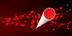
Vladimir Zharov's team of researchers has discovered a way to capture tumor cells in the bloodstream.

Nov. 17, 2009
A team led by University of Arkansas for Medical Sciences (UAMS) researchers on the cutting edge of nanotechnology has found a way to capture tumor cells in the bloodstream that could dramatically improve earlier cancer diagnosis and prevent deadly metastasis.
The discovery was published Nov. 15 in Nature Nanotechnology, a prestigious monthly print and online journal that provides a forum for leading research papers in all areas of nanoscience and nanotechnology. To read the abstract, click here.
Vladimir Zharov, director of the Phillips Classic Laser and Nanomedicine Laboratory at UAMS, said his team of researchers can inject a cocktail of magnetic and gold nanoparticles with a special biological coating into the bloodstream to target circulating tumor cells. A magnet attached to the skin above peripheral blood vessels can then capture the cells.
“By magnetically collecting most of the tumor cells from blood circulating in vessels throughout the whole body, this new method can potentially increase specificity and sensitivity up to 1,000 times compared to existing technology,” Zharov said.
Once the tumor cells are targeted and captured by the magnet, they can either be microsurgically removed from vessels for further genetic analysis or can be noninvasively eradicated directly in blood vessels by laser irradiation through the skin that is still safe for normal blood cells.
Zharov’s team, which has recently been awarded more than $3.7 million in clinical nanomedicine-related grants, includes Ekaterina Galanzha, M.D., Ph.D., an assistant professor in the UAMS Department of Otolaryngology; Evgeny Shashkov, Ph.D., a visiting scholar and laser physicist; Thomas Kelly, Ph.D., associate professor in the UAMS Department of Pathology; Jin-Woo Kim, Ph.D., a nano-biotechnologist at the University of Arkansas at Fayetteville; and Lily Yang, Ph.D., a biologist from Emory University.
A second related discovery by Zharov’s team was published in Cancer Research in October. It demonstrated that periodic laser irradiation of blood vessels decreases the level of circulating metastatic tumor cells more than 10 times and eventually led to an interruption of metastasis development in distant organs. To read the abstract, click here.
“Further study could determine whether these new cancer treatments are effective enough to be used alone or if they should be used in conjunction with conventional cancer therapy,” Zharov said.
The discovery highlighted in Cancer Research earned Zharov and his team a selection for Faculty of 1000 Biology, an award-winning Web site that highlights and evaluates the most interesting papers published in the biological sciences. Papers are selected based on the recommendations of more than 2000 of the world’s top researchers.
The new discoveries can also be applied for early detection of cancer recurrence and for real-time monitoring therapy efficiency involving the counting of circulating tumor cells.
“Most cancer deaths are the result of metastasis due to the spread of tumor cells from the primary tumor through the blood,” said James Suen, M.D., chairman of the UAMS Winthrop P. Rockefeller Cancer Institute’s Department of Otolaryngology, Head and Neck Surgery. “This revolutionary discovery introduced by Zharov’s team gives many patients hope in earlier cancer diagnosis and better treatment. The nanomedicine-based approach to read and treat whole blood in the body with nanotechnology seems to be universal, with further development holding the promise for the diagnosis and treatment of many diseases, including infections or cardiovascular disorders to prevent stroke and heart attack.”
UAMS is the state’s only comprehensive academic health center, with five colleges, a graduate school, a new 540,000-square-foot hospital, six centers of excellence and a statewide network of regional centers. UAMS has 2,775 students and 748 medical residents. Its centers of excellence include the Winthrop P. Rockefeller Cancer Institute, the Jackson T. Stephens Spine & Neurosciences Institute, the Myeloma Institute for Research and Therapy, the Harvey & Bernice Jones Eye Institute, the Psychiatric Research Institute and the Donald W. Reynolds Institute on Aging. It is the state’s largest public employer with more than 10,000 employees, including nearly 1,150 physicians who provide medical care to patients at UAMS, Arkansas Children’s Hospital, the VA Medical Center and UAMS’ Area Health Education Centers throughout the state. Visit www.uams.edu or www.uamshealth.com.Source
 Imagine nano-sized magnetic particles capable of fabulous feats such as killing cancer cells in the body, regenerating human tissue and skimming toxic oil spills from lakes and rivers.