Showing posts with label tumors. Show all posts

Tuesday, September 16, 2008

Researchers Develop Nano-Sized ‘Cargo Ships’ to Target and Destroy Tumors

September 11, 2008

By Kim McDonald

Scientists have developed nanometer-sized ‘cargo ships’ that can sail throughout the body via the bloodstream without immediate detection from the body’s immune radar system and ferry their cargo of anti-cancer drugs and markers into tumors that might otherwise go untreated or undetected.

Photo of Ji-Ho Park holding a vial containing the nanometer-sized cargo ships
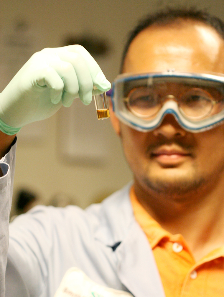
UCSD graduate student Ji-Ho Park holds a vial containing the nanometer-sized cargo ships, composed of a magnetic nanoparticle, a fluorescent quantum dot and an anti-cancer drug molecule that will be left on the site of the tumor.

Credit: Luo Gu, UCSD

In a forthcoming issue of the Germany-based chemistry journal Angewandte Chemie, scientists at UC San Diego, UC Santa Barbara and MIT report that their nano-cargo-ship system integrates therapeutic and diagnostic functions into a single device that avoids rapid removal by the body’s natural immune system. Their paper is now accessible in an early online version here.

“The idea involves encapsulating imaging agents and drugs into a protective ‘mother ship’ that evades the natural processes that normally would remove these payloads if they were unprotected,” said Michael Sailor, a professor of chemistry and biochemistry at UCSD who headed the team of chemists, biologists and engineers that turned the fanciful concept into reality. “These mother ships are only 50 nanometers in diameter, or 1,000 times smaller than the diameter of a human hair, and are equipped with an array of molecules on their surfaces that enable them to find and penetrate tumor cells in the body.”

These microscopic cargo ships could one day provide the means to more effectively deliver toxic anti-cancer drugs to tumors in high concentrations without negatively impacting other parts of the body.

“Many drugs look promising in the laboratory, but fail in humans because they do not reach the diseased tissue in time or at concentrations high enough to be effective,” said Sangeeta Bhatia, a physician, bioengineer and professor of Health Sciences and Technology at MIT who played a key role in the development. “These drugs don’t have the capability to avoid the body’s natural defenses or to discriminate their intended targets from healthy tissues. In addition, we lack the tools to detect diseases such as cancer at the earliest stages of development, when therapies can be most effective.”

The researchers designed the hull of the ships to evade detection by constructing them of specially modified lipids--a primary component of the surface of natural cells. The lipids were modified in such a way as to enable them to circulate in the bloodstream for many hours before being eliminated. This was demonstrated by the researchers in a series of experiments with mice.

The researchers also designed the material of the hull to be strong enough to prevent accidental release of its cargo while circulating through the bloodstream. Tethered to the surface of the hull is a protein called F3, a molecule that sticks to cancer cells. Prepared in the laboratory of Erkki Ruoslahti, a cell biologist and professor at the Burnham Institute for Medical Research at UC Santa Barbara, F3 was engineered to specifically home in on tumor cell surfaces and then transport itself into their nuclei.

Photo of a vial of anti-cancer nano ships glows red under a black light.
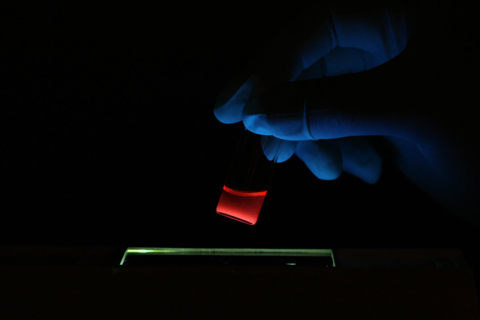
A vial of anti-cancer nano ships glows red under a black light. The particles glow red because they contain fluorescent "quantum dot" nanoparticles.

Credit: Luo Gu, UCSD

“We are now constructing the next generation of smart tumor-targeting nanodevices,” said Ruoslahti. “We hope that these devices will improve the diagnostic imaging of cancer and allow pinpoint targeting of treatments into cancerous tumors.”

The researchers loaded their ships with three payloads before injecting them in the mice. Two types of nanoparticles, superparamagnetic iron oxide and fluorescent quantum dots, were placed in the ship’s cargo hold, along with the anti-cancer drug doxorubicin. The iron oxide nanoparticles allow the ships to show up in a Magnetic Resonance Imaging, or MRI, scan, while the quantum dots can be seen with another type of imaging tool, a fluorescence scanner.

“The fluorescence image provides higher resolution than MRI,” said Sailor. “One can imagine a surgeon identifying the specific location of a tumor in the body before surgery with an MRI scan, then using fluorescence imaging to find and remove all parts of the tumor during the operation.”

The team found to its surprise in its experiments that a single mother-ship can carry multiple iron oxide nanoparticles, which increases their brightness in the MRI image.

“The ability of these nanostructures to carry more than one superparamagnetic nanoparticle makes them easier to see by MRI, which should translate to earlier detection of smaller tumors,” said Sailor. “The fact that the ships can carry very dissimilar payloads—a magnetic nanoparticle, a fluorescent quantum dot, and a small molecule drug—was a real surprise.”

The researchers noted that the construction of so-called “hybrid nanosystems” that contain multiple different types of nanoparticles is being explored by several other research groups. While hybrids have been used for various laboratory applications outside of living systems, said Sailor, there are limited studies done in vivo, or within live organisms, particularly for cancer imaging and therapy.

“That’s because of the poor stability and short circulation times within the blood generally observed for these more complicated nanostructures,” he added. As a result, the latest study is unique in one important way.

Illustration of a vial of anti-cancer nano ships glows red under a black light.
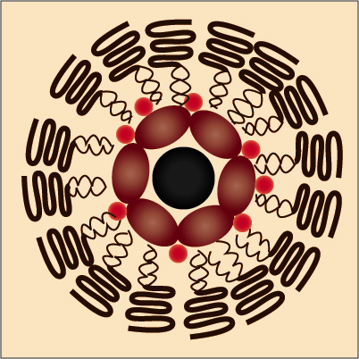
The nanometer-sized cargo ships look individually like a chocolate-covered nut cluster, in which a biocompatible lipid forms the chocolate shell and magnetic nanoparticles, quantum dots and the drug doxorubicin are the nuts.

Credit: Ji-Ho Park, UCSD

“This study provides the first example of a single nanomaterial used for simultaneous drug delivery and multimode imaging of diseased tissue in a live animal,” said Ji-Ho Park, a graduate student in Sailor’s laboratory who was part of the team. Geoffrey von Maltzahn, a graduate student working in Bhatia’s laboratory, was also involved in the project, which was financed by a grant from the National Cancer Institute of the National Institutes of Health.

The nano mother ships look individually like a chocolate-covered nut cluster, in which a biocompatible lipid forms the chocolate shell and magnetic nanoparticles, quantum dots and the drug doxorubicin are the nuts. They sail through the bloodstream in groups that, under the electron microscope, look like small, broken strands of pearls.

The researchers are now working on developing ways to chemically treat the exteriors of the nano ships with specific chemical “zip codes,” that will allow them to be delivered to specific tumors, organs and other sites in the body.

Media Contact: Kim McDonald, 858-534-7572
Comment: Michael Sailor, 858-534-8188

Source

Tuesday, July 29, 2008

New disease-fighting nanoparticles look like miniature pastries

July 29, 2008
By Gwen Ericson

-- Ultra-miniature bialy-shaped particles — called nanobialys because they resemble tiny versions of the flat, onion-topped rolls popular in New York City — could soon be carrying medicinal compounds through patients' bloodstreams to tumors or atherosclerotic plaques.

The nanobialys are an important addition to the stock of diagnostic and disease-fighting nanoparticles developed by researchers in the Consortium for Translational Research in Advanced Imaging and Nanomedicine (C-TRAIN) at Washington University School of Medicine in St. Louis. C-TRAIN's "smart" nanoparticles can deliver drugs and imaging agents directly to the site of tumors and plaques.

The new nanobialys weren't cooked up for their appealing shape — that's a natural result of the manufacturing process. The nanobialys answered a need for an alternative to the research group's gadolinium-containing nanoparticles, which were created for their high visibility in magnetic resonance imaging (MRI) scans.

Gadolinium is a common contrast agent for MRI scans, but recent studies have shown that it can be harmful to some patients with severe kidney disease.

"The nanobialys contain manganese instead of gadolinium," says first author Dipanjan Pan, Ph.D., research instructor in medicine in the Cardiovascular Division. "Manganese is an element found naturally in the body. In addition, the manganese in the nanobialys is tied up so it stays with the particles, making them very safe."

The bulk of a nanobialy is a synthetic polymer that can accept a variety of medical, imaging or targeting components. In the July 2008 issue of the Journal of the American Chemical Society the researchers report that targeted manganese-carrying nanobialys readily attached themselves to fibrin molecules, which are found in atherosclerotic plaques and blood clots. Laboratory-made clots then glowed brightly in MRI scans. They also showed that the nanobialys could carry both water-soluble and insoluble drugs.

Pan, who is a research instructor in medicine, played a leading role in the creation of nanobialys and chose the particles' name. "When we looked at the particles with an electron microscope, we saw they are round and flat, with a dimple in the center, like red blood cells, but also a little irregular, like bagels," he says. "I came across the word bialy, which is a Polish roll like a bagel without a hole that can be made with different toppings. So I called the particles nanobialys."

Pan is one of a group of researchers headed by Gregory M. Lanza, M.D., Ph.D., and Samuel A. Wickline, M.D. Lanza is an associate professor of medicine and biomedical engineering. Wickline is a professor of medicine, physics, biomedical engineering and cell biology and physiology. Lanza and Wickline are Washington University cardiologists at Barnes-Jewish Hospital.

Nanoparticles can be a more effective way to administer medications and imaging contrast agents because they are targeted, packaged units — drugs and imaging agents stay on the nanoparticles, which can be made to concentrate at a specific site in the body.

In animal studies, the research group has shown that their original, spherical nanoparticles can carry therapeutic compounds to tumors and atherosclerotic plaques. These nanoparticles also can hold thousands of molecules of gadolinium, which allows the researchers to use standard MRI scanning equipment to see where the nanoparticles congregate. The scans can then detect the size of lesions as well as the effect of drugs delivered by the nanoparticles.

But gadolinium has recently been linked to nephrogenic systemic fibrosis (NSF). First described in 2000, NSF is an unusual progressive, incurable disease seen in about 3 percent of patients with severe kidney disease who have had MRI scans using gadolinium. In NSF, collagen accumulates in tissues causing skin hardening and thickening, joint stiffening that can lead to physical disability, and disorders of the liver, lungs, muscles and heart.

"Even though it seems that gadolinium affects only those with severe renal failure, physicians have decided not to use gadolinium even in those with moderate renal failure," Lanza says. "A lot of patients with diabetes or hypertension develop renal failure, so that decision potentially affects many people. Our goal has always been that our nanoparticle technology should be able to help everyone. And with a growing number of people having diabetes and related cardiovascular problems, we knew we needed to find a substitute for gadolinium-based particles — nanobialys are our first step in that direction."

The researchers will continue to adapt the nanobialys for a variety of medicinal applications and work to develop other types of nanoparticles so that they can supply a wide range of medical needs.

"We're not sitting in the lab generating nanoparticles and then looking for what they could be used for," Lanza says. "We see a medical problem and ask what kind of particle might overcome it and then try to create it."


Pan D, Caruthers SD, Hu G, Senpan A, Scott MJ, Gaffney PJ, Wickline SA, Lanza GM. Ligand-directed nanobialys as theranostic agent for drug delivery and manganese-based magnetic resonance imaging of vascular targets. Journal of the American Chemical Society 2008 Jul 23;130(29):9186-7.

Source

Wednesday, May 7, 2008

Researchers Target Tumors With Tiny 'Nanoworms'

ScienceDaily (May 7, 2008) — Scientists at UC San Diego, UC Santa Barbara and MIT have developed nanometer-sized “nanoworms” that can cruise through the bloodstream without significant interference from the body’s immune defense system and—like tiny anti-cancer missiles—home in on tumors.

Their discovery, detailed in this week’s issue of the journal Advanced Materials, is reminiscent of the 1966 science fiction movie, the Fantastic Voyage, in which a submarine is shrunken to microscopic dimensions, then injected into the bloodstream to remove a blood clot from a diplomat’s brain.

Using nanoworms, doctors should eventually be able to target and reveal the location of developing tumors that are too small to detect by conventional methods. Carrying payloads targeted to specific features on tumors, these microscopic vehicles could also one day provide the means to more effectively deliver toxic anti-cancer drugs to these tumors in high concentrations without negatively impacting other parts of the body.

“Most nanoparticles are recognized by the body's protective mechanisms, which capture and remove them from the bloodstream within a few minutes,” said Michael Sailor, a professor of chemistry and biochemistry at UC San Diego who headed the research team. “The reason these worms work so well is due to a combination of their shape and to a polymer coating on their surfaces that allows the nanoworms to evade these natural elimination processes. As a result, our nanoworms can circulate in the body of a mouse for many hours.”

“When attached to drugs, these nanoworms could offer physicians the ability to increase the efficacy of drugs by allowing them to deliver them directly to the tumors,” said Sangeeta Bhatia, a physician, bioengineer and a professor of Health Sciences and Technology at MIT who was part of the team. “They could decrease the side effects of toxic anti-cancer drugs by limiting their exposure of normal tissues and provide a better diagnosis of tumors and abnormal lymph nodes.”

The scientists constructed their nanoworms from spherical iron oxide nanoparticles that join together, like segments of an earthworm, to produce tiny gummy worm-like structures about 30 nanometers long—or about 3 million times smaller than an earthworm. Their iron-oxide composition allows the nanoworms to show up brightly in diagnostic devices, specifically the MRI, or magnetic resonance imaging, machines that are used to find tumors.

“The iron oxide used in the nanoworms has a property of superparamagnetism, which makes them show up very brightly in MRI,” said Sailor. “The magnetism of the individual iron oxide segments, typically eight per nanoworm, combine to provide a much larger signal than can be observed if the segments are separated. This translates to a better ability to see smaller tumors, hopefully enabling physicians to make their diagnosis of cancer at earlier stages of development.”

In addition to the polymer coating, which is derived from the biopolymer dextran, the scientists coated their nanoworms with a tumor-specific targeting molecule, a peptide called F3, developed in the laboratory of Erkki Ruoslahti, a cell biologist and professor at the Burnham Institute for Medical Research at UC Santa Barbara. This peptide allows the nanoworms to target and home in on tumors.

“Because of its elongated shape, the nanoworm can carry many F3 molecules that can simultaneously bind to the tumor surface,” said Sailor. “And this cooperative effect significantly improves the ability of the nanoworm to attach to a tumor.”

The scientists were able to verify in their experiments that their nanoworms homed in on tumor sites by injecting them into the bloodstream of mice with tumors and following the aggregation of the nanoworms on the tumors. They found that the nanoworms, unlike the spherical nanoparticles of similar size that were shuttled out of the blood by the immune system, remained in the bloodstream for hours.

“This is an important property because the longer these nanoworms can stay in the bloodstream, the more chances they have to hit their targets, the tumors,” said Ji-Ho Park, a UC San Diego graduate student in materials science and engineering working in Sailor’s laboratory.

Park was the motivating force behind the discovery when he found by accident that the gummy worm aggregates of nanoparticles stayed for hours in the bloodstream despite their relatively large size.

While it’s not clear yet to the researchers why, Park notes that “the nanoworm’s flexibly moving, one dimensional structure may be one the reasons for its long life in the bloodstream.”

The researchers are now working on developing ways to attach drugs to the nanoworms and chemically treating their exteriors with specific chemical “zip codes,” that will allow them to be delivered to specific tumors, organs and other sites in the body.

“We are now using nanoworms to construct the next generation of smart tumor-targeting nanodevices,” said Ruoslahti. We hope that these devices will improve the diagnostic imaging of cancer and allow pinpoint targeting of treatments into cancerous tumors.”

Other researchers involved in the development were Michael Schwartz of UC San Diego, Geoffrey von Maltzahn of MIT, and Lianglin Zhang of UC Santa Barbara. The project was funded by grants from the National Cancer Institute of the National Institutes of Health.

Adapted from materials provided by University of California, San Diego. Original article written by Kim McDonald.